-
|
-
CX3CR1 (C-X3-C motif chemokine receptor 1), also known as fractalkine/CX3CL1 receptor, is a transmembrane protein expressed on the surface of immune cells that functions like an internal GPS receiver. Without it, the immune system would be driving blind, unable to navigate toward the distress signals coming from damaged tissues. As the sole high-affinity receptor for CX3CL1, CX3CR1 translates ligand binding into signaling cascades that remodel the cytoskeleton, enforce adhesion, and drive directional migration. In short, the CX3CL1–CX3CR1 axis ensures that defense cells not only know something is wrong, but also know exactly where to go and what to do (Imai et al., 1997).
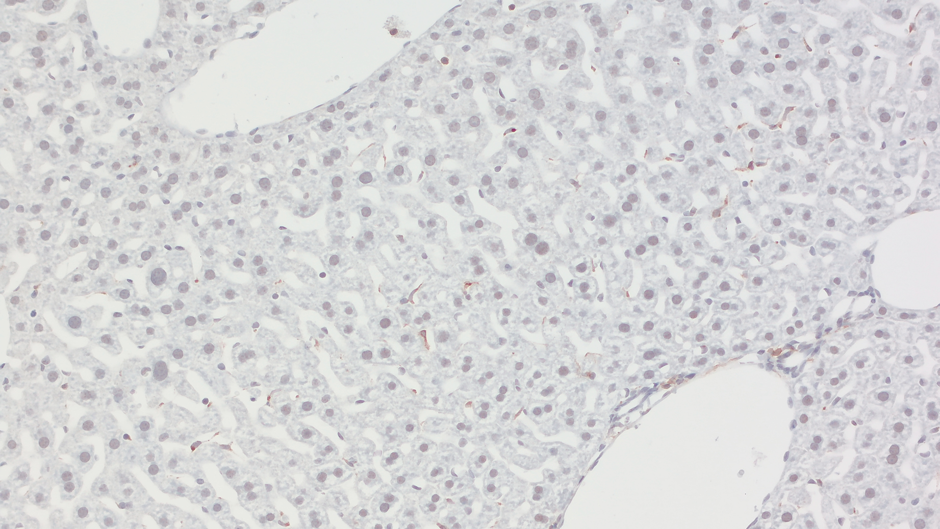
Figure 1: Detection of CX3CR1-positive Kupffer cells in a formalin fixed paraffin embedded (FFPE) mouse liver section with rabbit anti-CX3CR1 antibody (cat. no. HS-516 003, dilution 1:100, DAB, brown). Nuclei have been visualized by hematoxylin staining (blue).
CX3CR1 is a 7-transmembrane receptor coupled to heterotrimeric G proteins (GPCRs). Its ligand CX3CL1 is unusual among chemokines because it exists in both a membrane-bound and a soluble form, which is cleaved from the native transmembrane form by metalloproteinases (MMPs). The soluble form has chemotactic properties and strongly attracts T-cells and monocytes. The membrane-bound form facilitates diapedesis and is responsible for cell-to-cell adhesion, especially by promoting the strong adhesion of leukocytes (monocytes) to activated endothelial cells with the subsequent formation of an extracellular matrix and angiogenesis (Szukiewicz, 2024). As a result, the CX3CL1-CX3CR1 axis participates both in adhesive interactions and chemotactic signaling.
CX3CR1 is widely used as a marker for microglia, monocytes, macrophages, and related myeloid cell populations. Aside from the prominent expression in the mononuclear myeloid compartment (Jung et al., 2000), CX3CR1 receptor expression has also been reported for a natural killer (NK) cell subset and certain T-cell populations (Imai et al., 1997) that contribute to antigen sampling, cytokine production, and cytotoxic surveillance outside the central nervous system (Lee et al., 2018). Through the CX3CR1–CX3CL1 axis, these leukocytes are guided along CX3CL1 gradients, adhere to activated endothelium, and are recruited into sites of tissue injury or infection (Imai and Yasuda, 2016).
Dysregulation of the CX3CR1 pathway has been implicated in several inflammatory disorders, including atherosclerosis, inflammatory bowel disease, and multiple sclerosis (Mei et al., 2023). Increasing evidence further links the CX3CL1–CX3CR1 axis to cancer metastasis, particularly in tumors with a propensity to spread to bone, brain, and lung tissue (Szukiewicz, 2024).
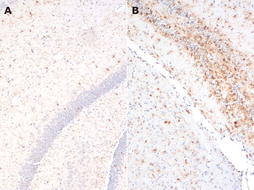
Figure 2: Indirect immunostaining of a formalin fixed paraffin embedded (FFPE) brain section from a mouse stroke model (middle cerebral artery occlusion (MCAO)) 14 days post-intervention with rabbit anti-CX3CR1 antibody (cat. no. HS-516 003, dilution 1:750, DAB, brown). CX3CR1 expression is higher in the ipsilateral hemisphere (B) compared with the contralateral hemisphere (A). Nuclei have been visualized by hematoxylin staining (blue).
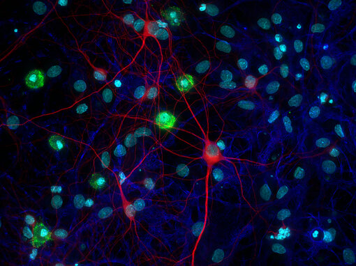
Figure 3: Indirect immunostaining of formaldehyde fixed rat cortex primary culture with rabbit anti-CX3CR1 antibody (cat. no. HS-516 003, dilution 1:500, green) for microglia, mouse anti-MAP2 antibody (cat. no. 188 011, dilution 1:1000, red) for neurons and chicken anti-GFAP antibody (cat. no. 173 006, dilution 1:1000, blue) for astrocytes. Nuclei have been visualized by DAPI staining (cyan).
Since some literature has specifically noted former limitations in the availability of anti-CX3CR1 antibodies, CX3CR1-EGFP reporter mice are widely used in microglia research (Jung et al., 2000, Schuster et al., 2012, Zhao et al., 2019).
In these mice, one of the CX3CR1 alleles has been replaced with an allele for endogenous GFP expression. But this experimental setup has its drawbacks, studies comparing EGFP with anti-CX3CR1 staining have found that EGFP fluorescence does not necessarily correlate with the amount of CX3CR1 protein on the cell surface (Meghraoui-Kheddar et al., 2020, Sommer et al., 2024).
To support different experimental strategies, the new SYSY Antibodies CX3CR1 product range includes the rabbit polyclonal antibody HS-516 003 and the rat monoclonal antibody HS-516 017. Together, these highly specific anti-CX3CR1 antibodies expand options for reliable CX3CR1 detection and offer a valuable alternative to CX3CR1-EGFP reporter mice, enabling detection of CX3CR1-positive cells in non-transgenic tissue and supporting flexible immunostaining and multiplex applications.
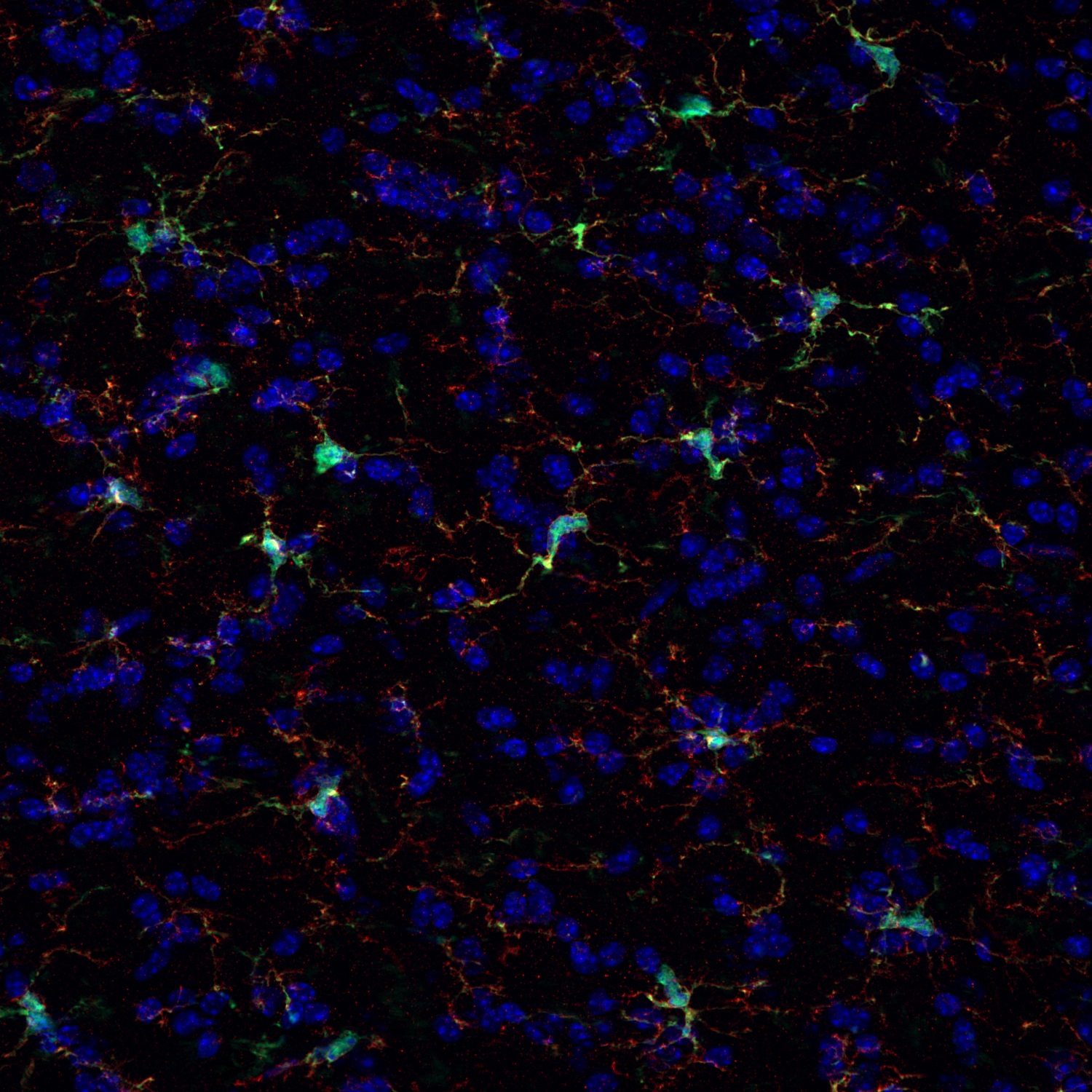
Figure 4: Indirect immunostaining of a formaldehyde fixed mouse brain section of a CX3CR1-EGFP transgene mouse with rat anti-CX3CR1 antibody (cat. no. 516 017, dilution 1:500, red) and a guinea pig anti-GFP (cat. no. 132 005, dilution 1:500, green). CX3CR1 antibody signal is colocalized with CX3CR1-EGFP. Nuclei have been visualized by DAPI staining (blue).
In the central nervous system, CX3CR1 is especially relevant for the analysis of microglia. Because of this, CX3CR1 is a relevant target in studies of neuroinflammation, neurodegeneration, brain development, and CNS homeostasis (Lee et al., 2010, Hoshiko et al., 2012).
In practice, however, CX3CR1 is often interpreted in combination with additional microglial markers to better distinguish resident microglia from infiltrating macrophages and other myeloid populations. These include TMEM119 and P2Y12, which are commonly regarded as relatively specific markers of homeostatic microglia, as well as Galectin-3, which is frequently associated with activated or disease-associated microglial states. Therefore, CX3CR1 is especially valuable when used as part of a broader marker panel for the phenotypic and functional characterization of microglia in health and disease (Bennett et al., 2016, Butovsky et al., 2014, Rai et al., 2020).
To further improve experimental analyses, CX3CR1 can also be combined with other lineage defining markers. Common markers paired with CX3CR1 include Ly6C for monocytes; F4/80, CD11b, and CD163 for macrophages; CD11c for dendritic cells; NK1.1 and/or NKp46 for NK cells.
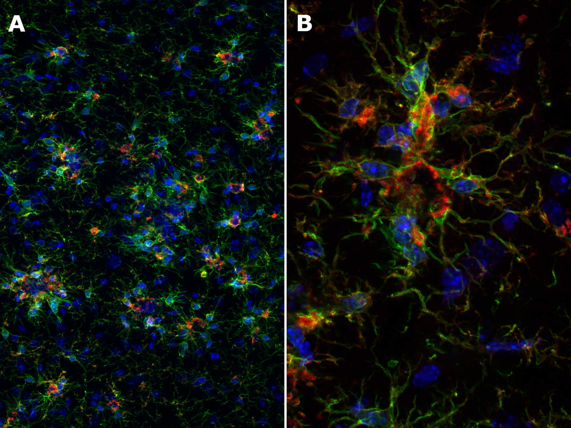
Figure 5: Indirect immunostaining of a formaldehyde-fixed mouse brain section with a rat anti-CX3CR1 antibody (cat. no. 516 017, 1:500, red) and a recombinant guinea pig anti-Iba1 antibody (cat. no. 234 308, 1:500, green), shown at 20× (A) and 63× (B) magnification. CX3CR1 expression is detected in Iba1-positive microglial cells. Nuclei were counterstained with DAPI (blue).
Find more information in our related Featured Topics
| Cat. No. | Product Description | Application | Quantity | Price | Cart |
|---|
| 384 117BT | CD11b, rat, monoclonal, purified IgG IgG, biotin mouse specific | WB IHC-P (FFPE) | 100 µg | $470.00 |   |
| HS-384 008 | CD11b, rabbit, monoclonal, recombinant IgGrecombinant IgG mouse specific | WB IHC IHC-P (FFPE) IHC-Fr | 100 µl | $420.00 | |
| HS-384 017 | CD11b, rat, monoclonal, purified IgG IgG human specific | WB IHC-P (FFPE) | 200 µl | $420.00 | |
| HS-384 018 | CD11b, rabbit, monoclonal, recombinant IgGrecombinant IgG human specific | WB IHC-P (FFPE) IHC-Fr | 100 µl | $420.00 | |
| HS-384 103 | CD11b, rabbit, polyclonal, affinity purifiedaffinity mouse specific | WB ICC IHC IHC-P (FFPE) | 200 µl | $455.00 | |
| HS-384 117 | CD11b, rat, monoclonal, purified IgG IgG mouse specific | WB IHC IHC-P (FFPE) IHC-Fr | 200 µl | $420.00 | |
| HS-384 308 | CD11b, Guinea pig, monoclonal, recombinant IgGrecombinant IgG mouse specific | WB IHC IHC-P (FFPE) IHC-Fr | 100 µl | $420.00 | |
| 375 004 | CD11c, Guinea pig, polyclonal, antiserumantiserum mouse specific | WB IHC IHC-P (FFPE) | 100 µl | $355.00 | |
| 375-0P | CD11c, control peptidecontrol peptide | 100 µg | $110.00 | | |
| HS-375 003 | CD11c, rabbit, polyclonal, affinity purifiedaffinity mouse specific | WB IHC IHC-P (FFPE) | 200 µl | $375.00 | |
| HS-375 008 | CD11c, rabbit, monoclonal, recombinant IgGrecombinant IgG mouse specific | IHC IHC-P (FFPE) | 50 µg | $420.00 | |
| HS-375 017 | CD11c, rat, monoclonal, purified IgG IgG mouse specific | IHC IHC-P (FFPE) IHC-Fr | 100 µg | $420.00 | |
| HS-493 017 | CD39, rat, monoclonal, purified IgG IgG mouse specific | WB IHC IHC-P (FFPE) | 100 µg | $420.00 | |
| HS-493 117 | CD39, rat, monoclonal, purified IgG IgG human specific | ICC IHC-P (FFPE) | 100 µg | $420.00 | |
| 427 117 | CD45, rat, monoclonal, purified IgG IgG human specific | IHC-P (FFPE) | 100 µg | $420.00 | |
Author: Dr. Roser Ufartes
Roser has a strong background in histopathology with special interest in understanding the origin and development of human diseases, especially the immune system and cancer. She is also part of the product developer team responsible for antibody development, validation and quality control.


Bennet et al., 2016: New tools for studying microglia in the mouse and human CNS. PMID: 26884166
Butovsky et al., 2014: Identification of a unique TGF-β-dependent molecular and functional signature in microglia. PMID: 24316888
Hoshiko et al., 2012: Deficiency of the microglial receptor CX3CR1 impairs postnatal functional development of thalamocortical synapses in the barrel cortex. PMID: 23100431
Imai et al., 1997: Identification and molecular characterization of fractalkine receptor CX3CR1, which mediates both leukocyte migration and adhesion. PMID: 9390561
Imai and Yasuda, 2016: Therapeutic intervention of inflammatory/immune diseases by inhibition of the fractalkine (CX3CL1)-CX3CR1 pathway. PMID: 29259682
Jung et al., 2000: Analysis of fractalkine receptor CX3CR1 function by targeted deletion and green fluorescent protein reporter gene insertion. PMID: 10805752
Lee et al., 2010: CX3CR1 deficiency alters microglial activation and reduces beta-amyloid deposition in two Alzheimer’s disease mouse models. PMID: 20864679
Lee et al., 2018: Tissue-specific Role of CX3CR1 Expressing Immune Cells and Their Relationships with Human Disease. PMID: 29503738
Meghraoui-Kheddar et al. 2020: Revising CX3CR1 Expression on Murine Classical and Non-classical Monocytes. PMID: 32582197
Mei et al., 2023: CX3CR1 expression predicts poor prognosis in paediatric acute myeloid leukaemia undergoing hyperleukocytosis. PMID: 36064206
Rai et al., 2020: Comparative analysis of human microglial models for studies of HIV replication and pathogenesis. PMID: 33213476
Schuster et al., 2012: Antibody validation by combining immunohistochemistry and protein extraction from formalin-fixed paraffin-embedded tissues. PMID: 22393911
Sommer et al., 2024: Discrepant Phenotyping of Monocytes Based on CX3CR1 and CCR2 Using Fluorescent Reporters and Antibodies. PMID: 38786041
Szukiewicz, 2024: CX3CL1 (Fractalkine)-CX3CR1 Axis in Inflammation-Induced Angiogenesis and Tumorigenesis. PMID: 38731899
Zhao et al., 2019: Targeting Microglia Using Cx3cr1-Cre Lines: Revisiting the Specificity. PMID: 31201215
Certificates
ISO 9001 2015 Quality Management System and Green Lab Platinum certification level for sustaining laboratory processes.


Newsletter
Sign up for our newsletter and get the latest updates and news.