NanoTag Biotechnologies and SYSY Antibodies introduce the first PSD95 FluoTag-X2 antibody that shows robust and reliable results in standard immunostaining applications like immunohistochemistry (IHC) (figure 1) and immunocytochemistry (ICC) (figure 2). It works on PFA / formaldehyde fixed tissues and cells from rat and mouse origin without any requirement for additional antigen retrieval (AGR) steps.
Figure 1: Indirect immunostaining of free floating PFA-fixed rat hippocampus (CA3) vibratome section with FluoTag-X2 anti-PSD95 Sulfo-Cyanine 3 (cat. no. N3702-SC3-L, dilution 1 : 500, red). Nuclei have been visualized by DAPI staining (blue).
Figure 2: Indirect immunostaining of PFA-fixed rat hippocampus neurons and astrocytes with FluoTag-X2 anti-PSD95-ATTO 542 (cat. no. N3702-At542-L, red) and FluoTag-X2 anti-GFAP-At488 (cat. no. N3802-At488-L, green). Nuclei have been visualized by DAPI staining (blue).
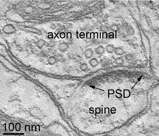
The post synaptic density (PSD) is a specialized protein dense region that localizes to the post-synaptic membrane juxtaposed to the pre-synaptic active zone. Originally, it has been identified by electron microscopy (EM) as an electron dense structure at post-synaptic membranes (figure 3). The PSD serves as a scaffolding structure at the synaptic cleft for the clustering of neurotransmitter receptors in close proximity to the pre-synaptic site of neurotransmitter release (Banker et al., 1974, Ziff, 1997, Dosemeci et al., 2016).
Depending on brain region, activity state, synapse maturation and in some neurodegenerative diseases the PSDs differ in size and composition (Meyer et al., 2014, Vyas and Montgomery, 2016).

Figure 3: EM tomography of a rat hippocampus synapse
N. Holderith PhD
Laboratory of Cellular Neurophysiology,
Institute of Experimental Medicine
Budapest
Figure 3: EM tomography of a rat hippocampus synapse
N. Holderith PhD
Laboratory of Cellular Neurophysiology,
Institute of Experimental Medicine
Budapest
PSD95 (postsynaptic density protein 95) also known as SAP90 (synapse associated protein 90) or Disks large homolog 4 is encoded by the DLG4 gene (Statthakis et al., 1997).
Together with its relatives PSD93, SAP102 and SAP97 it belongs to the MAGUK superfamily of proteins and shares the characteristic PDZ, SH3 and GUK domains (Woods and Bryant, 1993) (figure 4).
PSD95 and PSD93 are key components of the PSD (Hunt et al., 1996, Kennedy, 1997) and form multimeric scaffolds containing several hundred copies that cluster neurotransmitter receptors, ion-channels and other accompanying signaling proteins (Dosemeci et al., 2016).
For this reason PSD95 is a valuable and commonly used marker protein for the specific immunolabeling of post-synaptic sites in the nervous system both in immunocytochemistry (ICC) and immunohistochemistry (IHC). However, due to the multimeric, compact and dense structure antibody-epitopes are often masked or difficult to access by huge full IgG antibody molecules because of steric hindrance. Antigen retrieval methods or special adapted fixation protocols can improve the results but may be a restriction for the application of other antibodies in multiplexing experiments due to incompatibility with the additional tissue treatment steps.
The new PSD95 FluoTag N3702 is a single domain antibody (sdAb) frequently referred to as nanobody that has been developed by our partner company NanoTag Biotechnologies. This recombinant camelid derived antibody consists of only one antigen binding site of an alpaca heavy chain antibody. With only ~15 kDa, this FluoTag is about 10-times smaller than conventional IgG antibody molecules. Due to its small size this PSD95 FluoTag can reliably access its epitope even in standard 4% PFA perfusion-fixed and post-fixed tissue. This tissue preparation method is commonly used for the immunohistochemical staining of vibratome and cryo sections (free floating and slide mounted) since it ensures excellent preservation of tissue morphology and is compatible with a huge panel of available research antibodies.
Also in immunocytochemical staining of cultured neurons the newly introduced PSD95 FluoTag shows superior performance on PFA fixed cells (figure 2).
Its monovalent binding properties in combination with a defined number of fluorophore molecules per FluoTag make it an ideal reagent for quantitative analysis.
To ensure a maximum range of applications matching your needs, our new PSD95 FluoTag is available conjugated to a broad panel of fluorophores (see table).
| Cat. No. | Product Description | Application | Quantity | Price | Cart |
|---|
| N3702-AF568-L | PSD95 sdAb, camelid, monoclonal, FluoTag-X2FluoTag-X2, AZdye 568 | WB ICC IHC | 200 µl | $540.00 | |
| N3702-AF647-L | PSD95 sdAb, camelid, monoclonal, FluoTag-X2FluoTag-X2, Alexa 647 | ICC IHC IHC-P | 200 µl | $540.00 | |
| N3702-At488-L | PSD95 sdAb, camelid, monoclonal, FluoTag-X2FluoTag-X2, ATTO 488 | ICC IHC IHC-P | 200 µl | $540.00 |
Banker et al. 1974: Proteins of the postsynaptic density. PMID: 4419608
Dosemeci et al. 2016: The Postsynaptic Density: There Is More than Meets the Eye. PMID: 27594834
Hunt et al. 1996: PSD-95 is associated with the postsynaptic density and not with the presynaptic membrane at forebrain synapses. PMID: 8778289
Kennedy 1997: The postsynaptic density at glutamatergic synapses. PMID: 9185308
Meyer et al. 2014: Balance and stability of synaptic structures during synaptic plasticity. PMID: 24742464
Statthakis et al. 1997: Human postsynaptic density-95 (PSD95): location of the gene (DLG4) and possible function in nonneural as well as in neural tissues. PMID: 9286702
Vyas and Montgomery 2016: The role of postsynaptic density in neural degeneration and regeneration. PMID: 27482211
Woods and Bryant 1993: ZO-1, DlgA and PSD-95/SAP90: homologous proteins in tight, septate and synaptic cell junctions. PMID: 8155583
Ziff 1997: Enlightening the postsynaptic density. PMID: 9427241