-
|
-
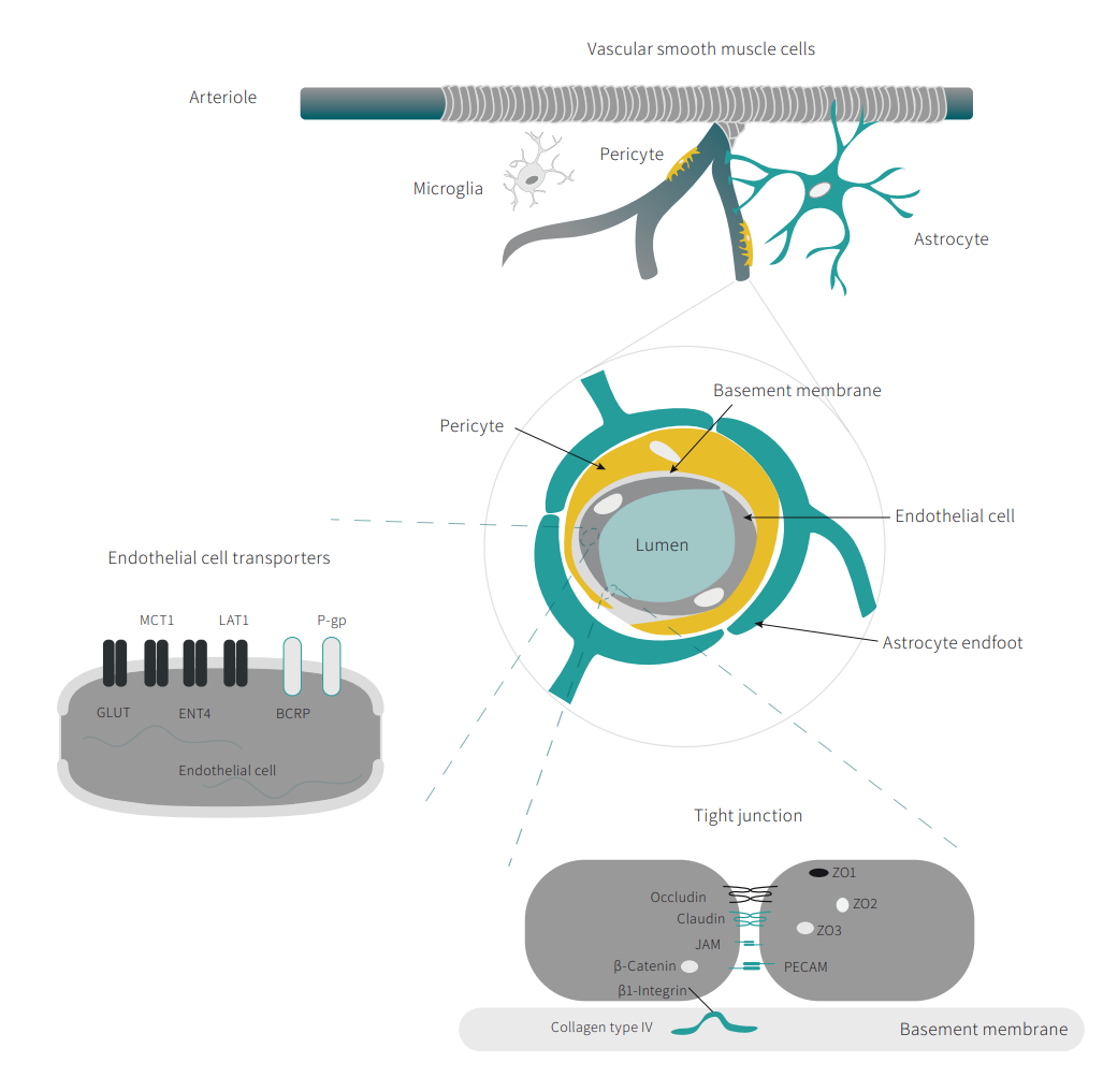
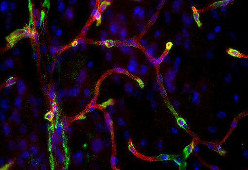
The blood-brain barrier (BBB) is a tightly regulated, highly selective interface that acts as a physical and metabolic barrier between the central nervous system (CNS) and the systemic circulation. Its primary function is to maintain CNS homeostasis by strictly controlling the movement of ions, nutrients, and cells into and out of the brain. This is achieved through a coordinated network of ion channels, transporters, and efflux pumps, as well as the structural and signaling contributions of vascular endothelial cells, mural cells (including pericytes and vascular smooth muscle cells (vSMCs)) and specialized cell-to-cell junctions. Disruption of the BBBs structural integrity has been linked to a variety of neurological disorders, such as neuroinflammation, neurodegeneration, and ischemic injury.
Find more information in our Featured Topic about Neuroinflammation.
The blood-brain barrier is primarily formed by brain microvascular endothelial cells, which are connected by complex tight junctions that restrict paracellular transport. These endothelial cells exhibit low rates of transcytosis and express specialized cell transporters that tightly regulate the movement of ions, nutrients, and metabolites between the blood and the brain.
The BBB is an essential component of the neurovascular unit (NVU), which also includes pericytes, astrocytes, neurons, and the extracellular matrix. Pericytes are embedded within the vascular basement membrane and contribute to vascular stability, angiogenesis, and BBB maintenance. Astrocytic endfeet tightly ensheath the cerebral vasculature and support endothelial cell function through the secretion of signaling molecules and metabolic factors. Together with the basement membrane, these cellular interactions maintain BBB integrity and CNS homeostasis (Galea, 2021, Kadry et al., 2020, Lochhead et al., 2020).
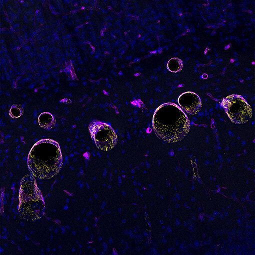
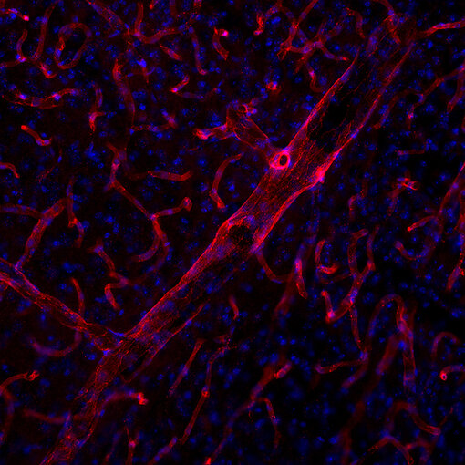
Endothelial cells (ECs) form the innermost cell layer of blood vessels. Their main function is to regulate the entry and exit of nutrients, ions, molecules, cells, and substances from the vascular system into and out of the surrounding tissue. ECs exhibit specialized features including a flat morphology, a lack of fenestrae, and reduced macropinocytosis and caveolar transcytosis (Galea, 2021). The inter-endothelial sealing is maintained by tight and adherens junctions that restrict both paracellular and transcellular transport (Abbott et al., 2010). Frequently used markers include Caveolin1, CD31, Mfsd2a and von Willebrand factor (vWF).
Figure 2: Indirect immunostaining of a formaldehyde fixed mouse brain (cortex) section with guinea pig anti-von Willebrand factor antibody (cat. no. 527 005, yellow) and rat anti-Collagen type IV antibody (cat. no. 462 017, magenta). Nuclei have been visualized by DAPI staining (blue). Antigen retrieval (10mM citrate, pH 6.0, overnight at 60°C) has been applied before staining.
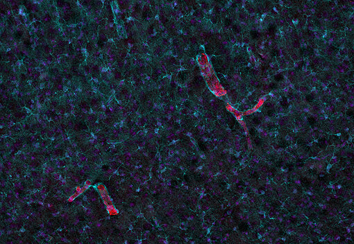
Figure 3: Indirect immunostaining of a formaldehyde fixed mouse brain section with rabbit anti-CD31 antibody (cat. no. HS-351 008, red). Nuclei have been visualized by DAPI staining (blue).
Molecular barrier functions are maintained by multiple classes of transporters localized to distinct luminal and abluminal membrane compartments. While small lipophilic molecules (typically <400 Da) and gases can diffuse across the endothelial membrane, water transport occurs both by passive diffusion and via aquaporin channels. The uptake of polar nutrients, ions, and larger biomolecules requires specialized mechanisms. Brain endothelial cells express a wide array of substrate-specific transporters and ion channels. Ion fluxes (Na+, K+, Cl-, HCO₃-, H+, Ca²+) are mediated by ATPases, uniporters, exchangers, and symporters. Carrier-mediated transport systems like GLUT1, MCT1 or LAT1 facilitate the uptake of glucose, amino acids, monocarboxylates, fatty acids, and other organic solutes. Larger macromolecules, such as insulin, transferrin, leptin, and apolipoprotein E-containing lipoproteins, enter via receptor-mediated transcytosis, whereas ABC efflux transporters (e.g., P-glycoprotein, BCRP, MRPs) remove xenobiotics and metabolic waste (Sweeney et al., 2019, Miller, 2016). Together with the inter-endothelial tight junctions, these transport pathways establish endothelial polarity, defining distinct luminal and abluminal membrane properties.
The endothelial cells (ECs) of the brain microvasculature are equipped with rows of dense regions of protein complexes, including tight and adherens junctions. These transmembrane protein complexes form a paracellular barrier between neighbouring microvascular endothelial cells of the brain and limit the free diffusion of substances through the paracellular space. Tight junctions (TJs) are predominantly located at the apical membrane (to the lumen), whereas adherens junctions (AJs) are positioned immediately underneath the TJs within the apical junctional complex. In endothelial cell-to-cell connections within the brain, both junctional complexes are also observed in a mixed configuration. TJs are made up of transmembrane protein complexes of three types of molecules: occludins, claudins, and intracellular junction adhesion molecules (JAMs) which are complemented by various accessory proteins of the cytoplasm such as zonula occludens-1, -2, -3 (ZO-1, ZO-2, ZO-3), cingulin, and others (Campbell et al., 2017, Galea, 2021, Kadry et al., 2020).
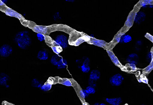
The basement membrane (BM) is a specialized extracellular matrix (ECM) that forms an integral component of the BBB. It is located between ECs, pericytes (PCs), and astrocytic endfeet and contributes to the structural organization of the neurovascular unit. In cerebral microvessels, the BM consists of two closely apposed layers: an endothelial BM, produced primarily by endothelial cells and pericytes, and a parenchymal BM, secreted by astrocytes and forming the vascular glia limitans.
Structurally, the BM is a thin, highly organized matrix approximately 50-100 nm thick. Its principal components include collagen IV, laminins, heparan sulfate proteoglycans, and nidogens, which together form a scaffold that provides mechanical stability and mediates cell-matrix interactions. In addition, the BM contains adhesion molecules and matrix-bound signaling factors that regulate cellular behavior.
Functionally, the BM serves as an important barrier to the movement of cells and macromolecules from the circulation into the brain parenchyma. Beyond its barrier role, the BM also contributes to several essential functions, including cell anchoring, signal transduction, and structural support (Thomsen et al., 2017, Xu et al., 2019, Galea, 2021).
Figure 8: Indirect immunostaining of a formaldehyde fixed rat brain section with guinea pig anti-Collagen IV antibody (cat. no. 462 004, white). Nuclei have been visualized by DAPI staining (blue). Antigen retrieval (0.2 mg/ml pepsin in 0.2 M HCl for 20 min at 37°C) has been applied before staining.
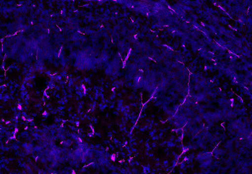
Figure 9: Indirect immunostaining of a formaldehyde fixed postnatal day 0 (P0) mouse brain section with guinea pig anti-Fibronectin antibody (cat. no. 550 005, purple). Nuclei have been visualized by DAPI staining (blue).
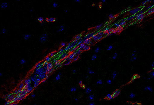
Mural cells include vessel-associated cell types such as pericytes and vascular smooth muscle cells (vSMCs). vSMCs encircle the endothelial layer and are densely arranged along the vessel. In larger vessels, they form concentric layers that provide structural stability. By mediating vasoconstriction and vasodilation, vSMCs are a critical component of the regulation of blood flow and vascular tone in the brain. vSMCs share markers with other mural cells (e.g., αSMA, PDGFRβ, desmin, CD13, Ng2, CD146), complicating classification. Differences in protein expression help distinguish mural cells, as vSMCs typically show high levels of αSMA, desmin, PDGFRβ, and CD13 but low NG2, whereas capillary pericytes show high PDGFRβ and CD13 but low desmin and αSMA levels, with expression patterns in the brain further varying depending on their vascular localization (Vanlandewijck et al., 2018, Smyth et al., 2018).
Pericytes dominate capillaries and post-capillary venules. They support BBB integrity by stabilizing vessels, regulating capillary diameter, and controlling local blood flow. Beyond mechanical functions, pericytes contribute to angiogenesis, matrix deposition, wound healing, and modulating vascular permeability, including immune cell infiltration (Brown et al., 2019, Kadry et al., 2020). The identification of pericytes remains challenging due to the lack of a unique molecular marker that can reliably distinguish them from other perivascular mural cells, such as vSMCs. In the murine brain, pericytes are typically identified by the expression of platelet-derived growth factor receptor beta (PDGFRβ) and the chondroitin sulfate proteoglycan NG2. Additional markers frequently used to characterize pericytes include alpha-smooth muscle actin (αSMA), desmin, CD146, CD13, and ATP13A5 (Attwell et al., 2016, Sweeney et al., 2016, Smyth et al., 2018).
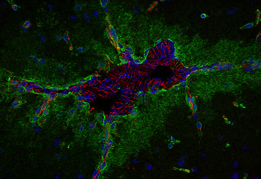
At the BBB, astrocytes provide a direct interface between the mural cells and the basement membrane of the vasculature through multiple processes. They also interact with neurons and other glial cells such as microglia and oligodendrocytes. Together, these cells form a functional unit known as the neurovascular unit (NVU) (Sweeney et al., 2018). Within the NVU, astrocytes establish a cellular connection between neuronal circuits and the cerebral blood flow. They coordinate vascular permeability, neuroimmune responses, and waste clearance in order to maintain the necessary homeostatic environment of the CNS (Abbott et al., 2006).
The contact point between astrocytes and the vasculature is formed by specialized subcellular foot-like terminal expansions, the astrocytic endfeet. Astrocyte endfeet enwrap the abluminal vessel side and form a gliovascular interface that covers almost 100% of the brain’s vasculature (Diaz-Castro et al., 2023, Mathiisen et al., 2010). Astrocyte endfeet are polarized structures that contain organelles, including a protein machinery, microtubules, bundles of intermediate filaments composed of glial fibrillary acidic protein (GFAP), and a variety of metabolic enzymes. The astrocyte-vasculature interaction is further mediated by the expression of channel proteins, transporters, adhesion and scaffold proteins (Abbott et al., 2006). Astrocytic endfeet proteins play a vital role in regulating water and ion homeostasis, especially for potassium, and facilitate waste removal. This function is primarily mediated by the high expression of Aquaporin4 (AQP4) water channels and the ATP-sensitive inward rectifier potassium channel Kir4.1 (Nagelhus and Ottersen, 2013).
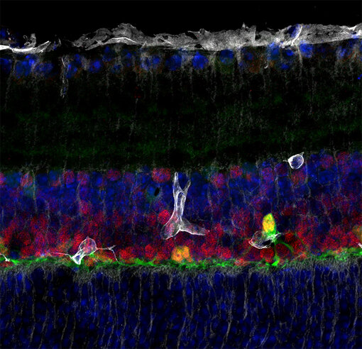
Figure 12: Indirect immunostaining of a formaldehyde fixed mouse retina section with mouse anti-Kir4.1 antibody (cat. no. 472 011, white), guinea pig anti-Prox1 antibody (cat. no. 509 005, red) and rabbit anti-Calbindin D28k antibody (cat. no. 214 008, green). Nuclei have been visualized by DAPI staining (blue). Antigen retrieval (10mM citrate, pH 6.0, overnight at 60°C) has been applied before staining.
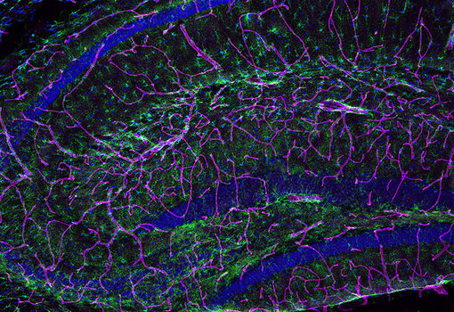
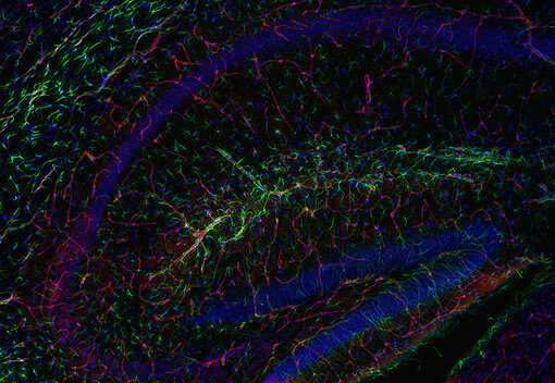
Figure 13: Indirect immunostaining of a formaldehyde fixed mouse hippocampus section with guinea pig anti-MLC1 antibody (cat. no. 525 005, red) and chicken anti-Aquaporin4 antibody (cat. no. 429 006, white). Nuclei have been visualized by DAPI staining (blue). Antigen retrieval (10mM citrate, pH 6.0, overnight at 60°C) has been applied before staining.
| Cat. No. | Product Description | Application | Quantity | Price | Cart |
|---|
| HS-511 008 | Desmin, rabbit, monoclonal, recombinant IgGrecombinant IgG | WB IHC IHC-P (FFPE) IHC-Fr | 50 µg | $420.00 |   |
| 369 003 | DOPA decarboxylase, rabbit, polyclonal, affinity purifiedaffinity | WB IHC IHC-P (FFPE) | 50 µg | $380.00 | |
| 369 004 | DOPA decarboxylase, Guinea pig, polyclonal, antiserumantiserum | IHC | 100 µl | $370.00 | |
| 369 008 | DOPA decarboxylase, rabbit, monoclonal, recombinant IgGrecombinant IgG | WB IHC IHC-P (FFPE) | 50 µg | $420.00 | |
| 369 011 | DOPA decarboxylase, mouse, monoclonal, purified IgG IgG | WB IHC IHC-P (FFPE) | 100 µg | $420.00 | |
| 369 104 | DOPA decarboxylase, Guinea pig, polyclonal, antiserumantiserum | IHC-P (FFPE) | 100 µl | $370.00 | |
| 497 003 | EGR1, rabbit, polyclonal, affinity purifiedaffinity | ICC IHC IHC-P (FFPE) | 50 µg | $380.00 | |
| 463 005 | ENT4, Guinea pig, polyclonal, affinity purifiedaffinity | WB ICC IHC | 50 µg | $465.00 | |
| 550 005 | Fibronectin, Guinea pig, polyclonal, affinity purifiedaffinity | WB ICC IHC IHC-P (FFPE) | 50 µg | $465.00 | |
| 419 005 | GLUT1, Guinea pig, polyclonal, affinity purifiedaffinity | IHC IHC-P (FFPE) IHC-Fr | 50 µg | $465.00 | |
| 522 004 | JAM-A, Guinea pig, polyclonal, antiserumantiserum | WB ICC | 100 µl | $370.00 | |
| 472 005 | Kir4.1, Guinea pig, polyclonal, affinity purifiedaffinity | WB IP ICC IHC IHC-P (FFPE) IHC-Fr | 50 µg | $465.00 | |
| 472 011 | Kir4.1, mouse, monoclonal, purified IgG IgG | WB IP ICC IHC IHC-P (FFPE) | 100 µg | $420.00 | |
| 519 005 | LAT1, Guinea pig, polyclonal, affinity purifiedaffinity | WB IHC-Fr IHC-G | 50 µg | $465.00 | |
| 517 003 | MCAM, rabbit, polyclonal, affinity purifiedaffinity | WB ICC IHC IHC-P (FFPE) IHC-Fr | 50 µg | $380.00 | |
Abbott NJ, Rönnbäck L, Hansson E. 2006 Astrocyte-endothelial interactions at the blood-brain barrier. PMID: 16371949
Abbott NJ, Patabendige AAK, Dolman DEM, Yusof SR, Begley DJ. 2010. Structure and function of the blood-brain barrier. PMID: 19664713
Attwell D, Mishra A et al. 2016 What is a pericyte? PMID: 26661200
Brown LS, Foster CG, Courtney JM et al. 2019. Pericytes and Neurovascular Function in the Healthy and Diseased Brain. PMID: 31316352
Campbell HK, Maiers JL, DeMali KA. 2017. Interplay Between Tight Junctions & Adherens Junctions. PMID: 28372972
Diaz-Castro B, Robel S, Mishra A. 2023 Astrocyte Endfeet in Brain Function and Pathology: Open Questions. PMID: 36854317
Galea I. 2021. The blood-brain barrier in systemic infection and inflammation. PMID: 34594000
Kadry H, Noorani B, Cucullo L. 2020. A blood-brain barrier overview on structure, function, impairment, and biomarkers of integrity. PMID: 33208141
Lochhead JJ, Yang J, Ronaldson PT, Davis TP. 2020. Structure, Function, and Regulation of the Blood-Brain Barrier Tight Junction in Central Nervous System Disorders. PMID: 32848858
Mathiisen TM, Lehre KP, Danbolt NC, Ottersen OP. 2010. The perivascular astroglial sheath provides a complete covering of the brain microvessels: an electron microscopic 3D reconstruction. PMID: 20468051
Miller DS, 2016. Regulation of ABC Transporters at the Blood-Brain Barrier. PMID: 25670036
Nagelhus EA and Ottersen OP. 2013. Physiological roles of aquaporin-4 in brain. PMID: 24137016
Smyth LCD et al. 2018. Markers for human brain pericytes and smooth muscle cells. PMID: 29885791
Sweeney MD, Zhao Z, Montagne A, Nelson AR, Zlokovic BV. 2019. Blood-Brain Barrier: From Physiology to Disease and Back. PMID: 30280653
Thomsen MS, Routhe LJ, Moos T. 2017. The vascular basement membrane in the healthy and pathological brain. PMID: 28753105
Vanlandewijck M et al. 2018. A molecular atlas of cell types and zonation in the brain vasculature. PMID: 29443965
Xu L, Nirwane A, Yao Y. 2019. Basement membrane and blood-brain barrier. PMID: 31338215
Certificates
ISO 9001 2015 Quality Management System and Green Lab Platinum certification level for sustaining laboratory processes.


Newsletter
Sign up for our newsletter and get the latest updates and news.