Cat. No.: HS-467 003
Amount: 50 µg
Price:
$375.00
|
|

|
|
| Cat. No. HS-467 003 |
50 µg specific antibody, lyophilized. Affinity purified with the immunogen. Albumin and azide were added for stabilization. For reconstitution add 50 µl H2O to get a 1mg/ml solution in PBS. Then aliquot and store at -20°C to -80°C until use. Antibodies should be stored at +4°C when still lyophilized. Do not freeze! |
| Applications | |
| Reactivity |
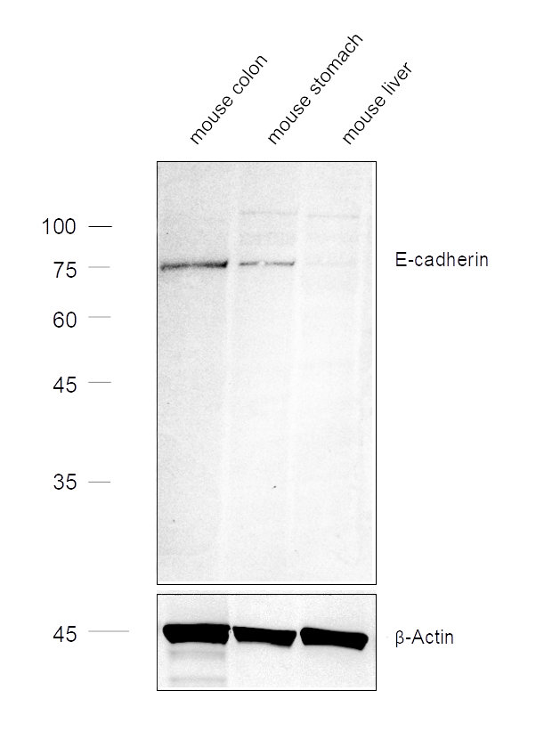
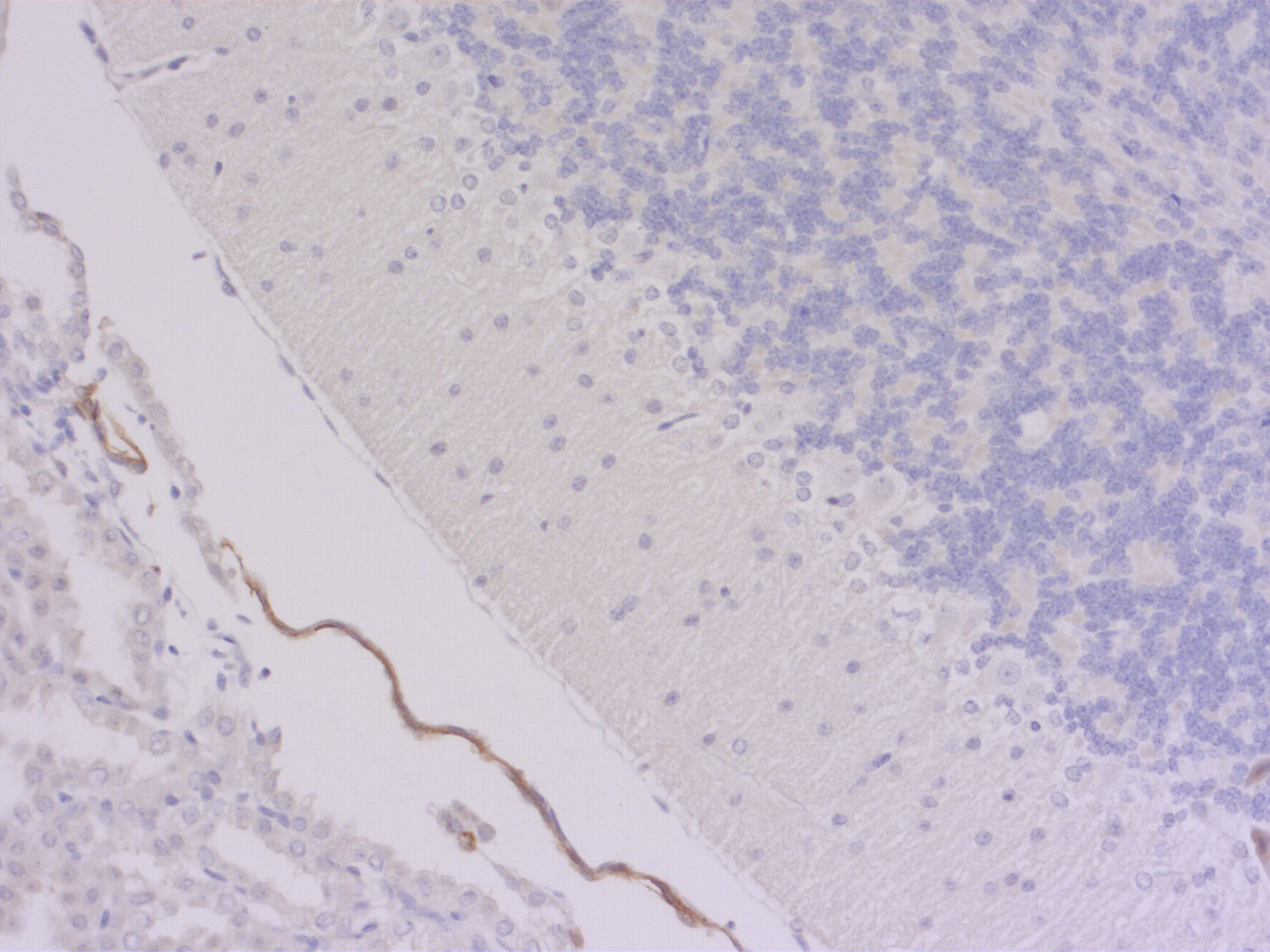
Reacts with: mouse (P09803), rat. No signal: human. Other species not tested yet. |
| Remarks |
IHC: Antigen retrieval with citrate buffer pH 6 is required. |
| Data sheet | Datasheet hs-467_003 |
|
|
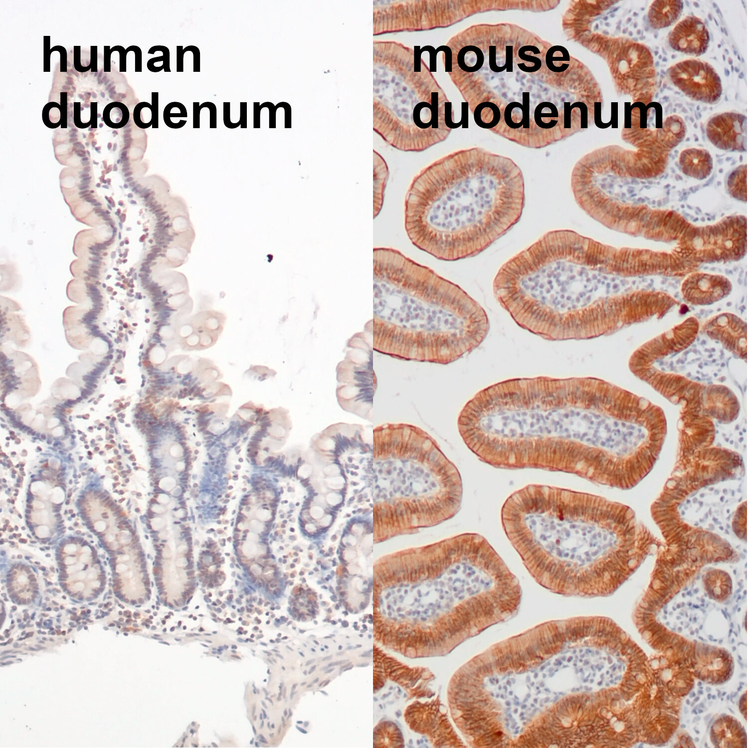
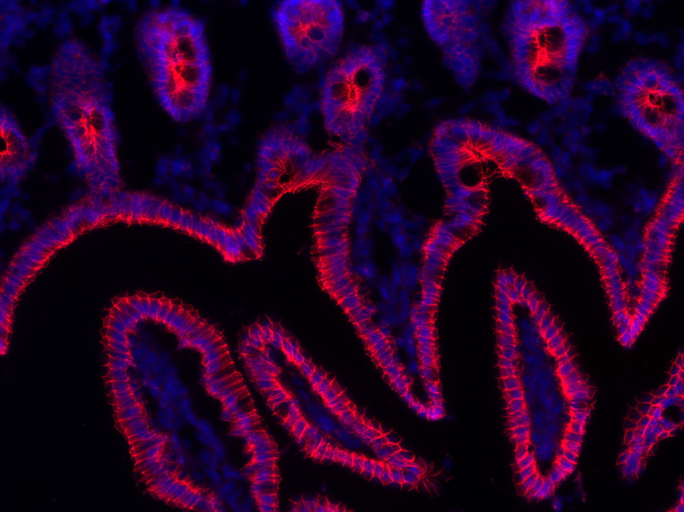
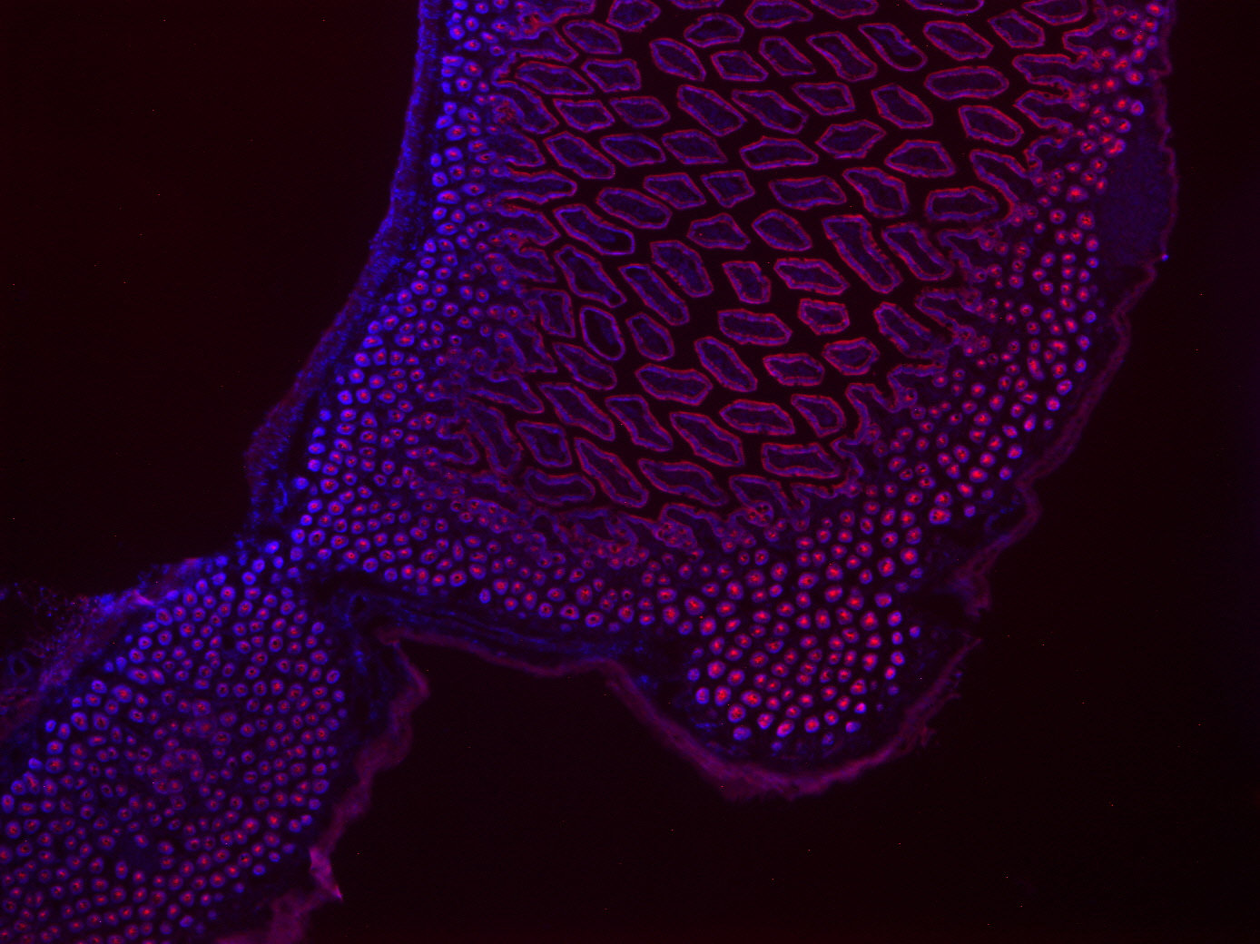
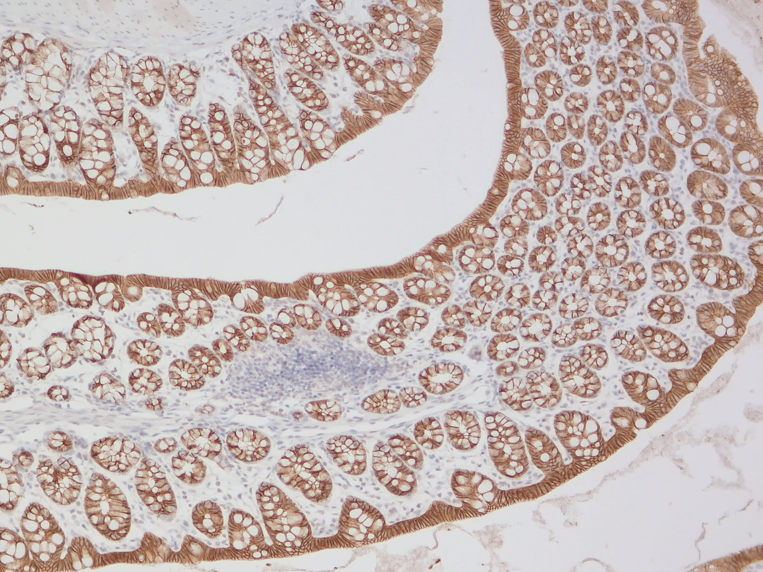
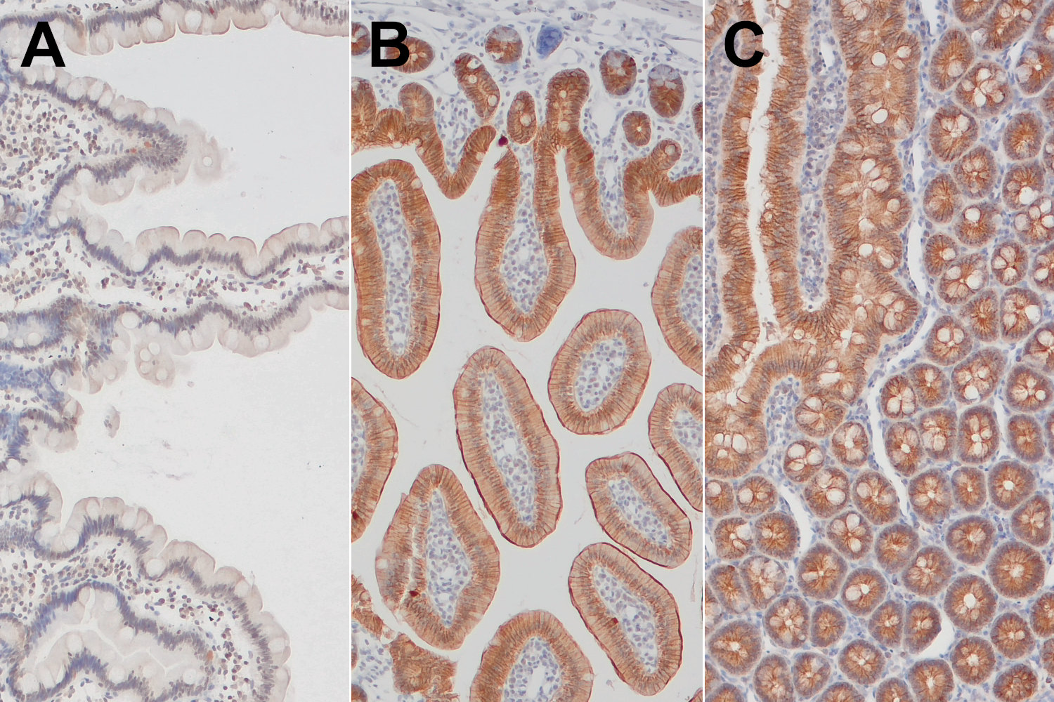
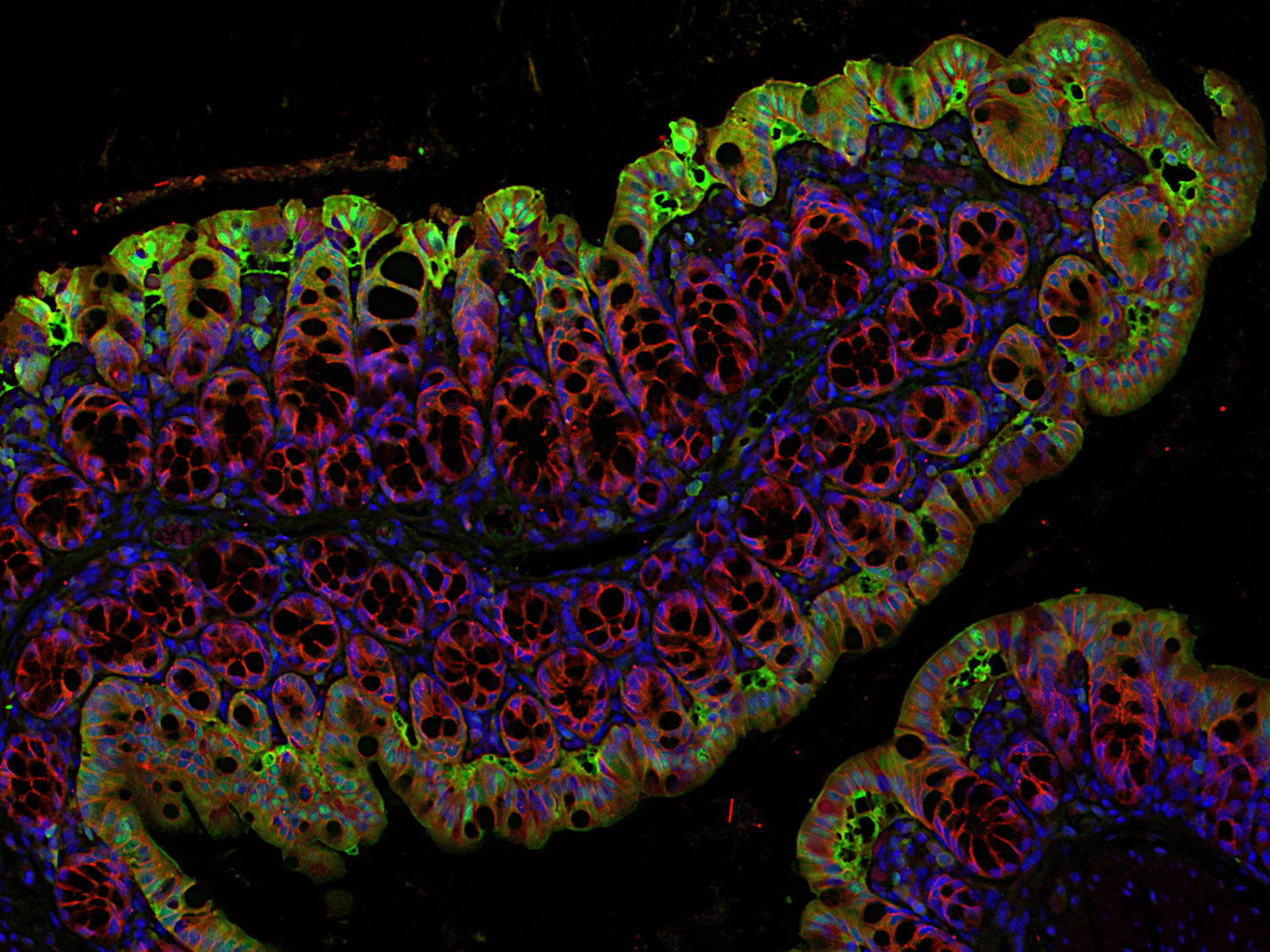
Indirect immunostaining of human and mouse duodenum sections using mouse-E-cadherin specific antibody.
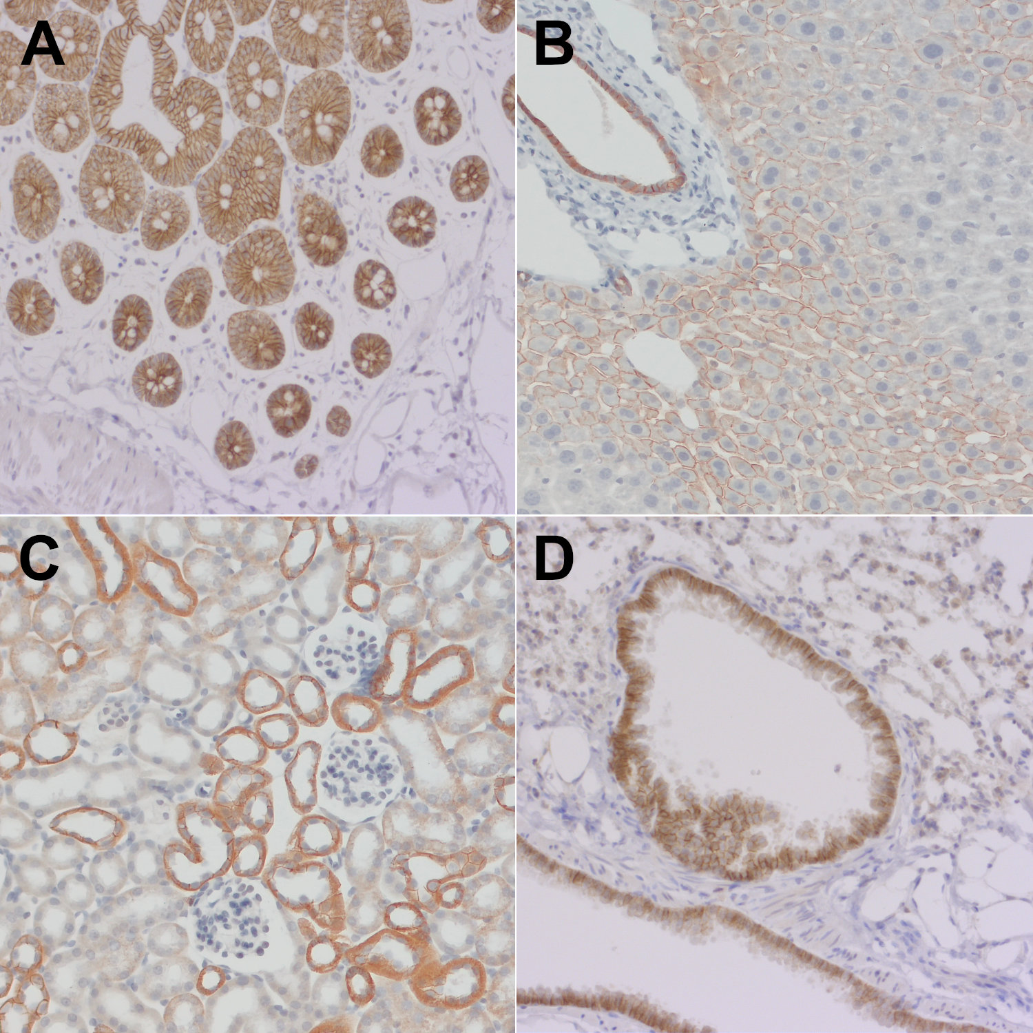
Epithelial cadherin (E-cadherin) also known as Cadherin-1, CAM 120/80 or uvomorulin belongs together with neuronal (N) cadherin to the type I classical cadherins, transmembrane proteins that function in calcium-dependent cell-cell adhesion (1). In normal tissues, E-cadherin is expressed by most epithelial cells. A distinct distribution of E-cadherin expression is found in the kidney, where only distal tubuli show E-cadherin expression and in the placenta, where only the cytotrophoblastic layer stains positive for E-cadherin (2). In the human normal adult nervous system E-cadherin expression is limited to the arachnoid membrane, whereas in mice E-cadherin is also expressed in neural stem cells, where E-cadherin regulates self-renewal (3). E-cadherin is a potent tumor suppressor and the so-called “cadherin switch” - downregulation of E-cadherin while N-cadherin is upregulated - is often found in malignant epithelial cancers. This Epithelial-to-Mesenchymal Transition (EMT) has been shown to be crucial in tumorigenesis where the EMT program enhances metastasis, chemoresistance and tumor stemness (4). However, also E-cadherin upregulation in malignancies derived from E-cadherin negative normal tissues tend to be linked to unfavorable tumor phenotype and disease outcome (2). In syngeneic mouse tumors E-cadherin expression is typically lower than in human tumors suggesting that syngeneic mouse models have a more mesenchymal-like tumor cellular phenotype (5).
Certificates
ISO 9001 2015 Quality Management System and Green Lab Platinum certification level for sustaining laboratory processes.


Newsletter
Sign up for our newsletter and get the latest updates and news.