Cat. No.: 404 017
Amount: 200 µl
Price:
$420.00
|
|

|
|
| Cat. No. 404 017 |
200 µl purified IgG, lyophilized. Albumin and azide were added for stabilization. For reconstitution add 200 µl H2O. Then aliquot and store at -20°C to -80°C until use. Antibodies should be stored at +4°C when still lyophilized. Do not freeze! |
| Applications | |
| Clone | 12G6 |
| Subtype | IgG2a (κ light chain) |
| Immunogen | Synthetic peptide corresponding to AA 8 to 19 from human LaminB1 (UniProt Id: P20700) |
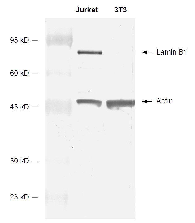
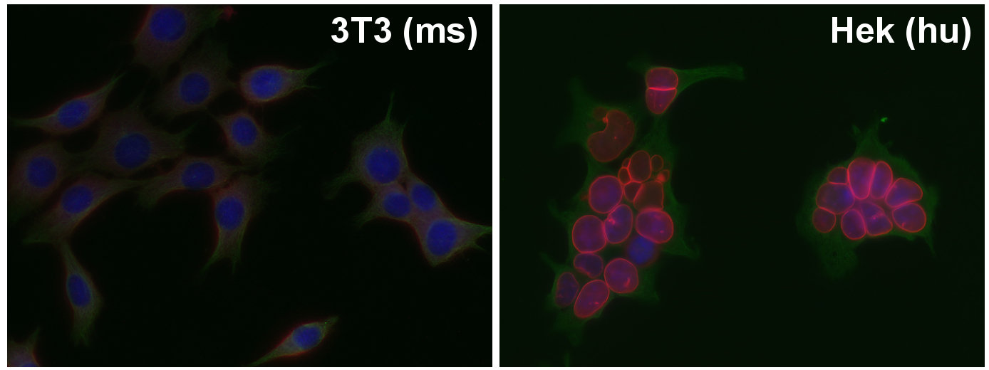
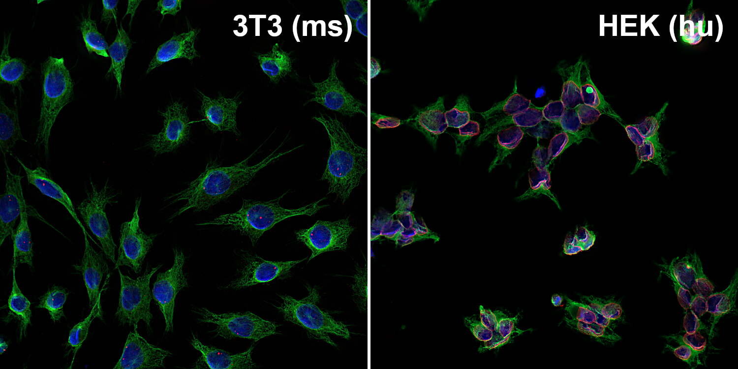
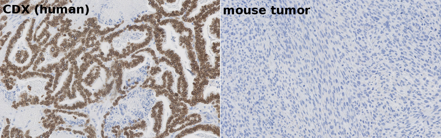
| Reactivity |
Reacts with: human (P20700), pig. No signal: mouse (P14733), rat (P70615). Other species not tested yet. |
| Remarks |
ICC: Methanol fixation is recommended. |
| Data sheet | Datasheet 404_017 |
|
|
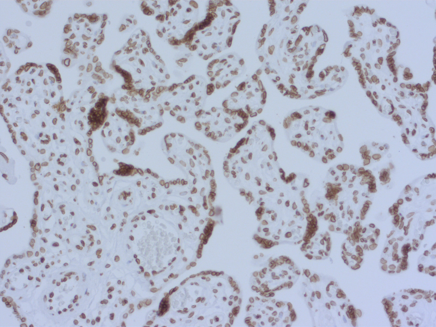
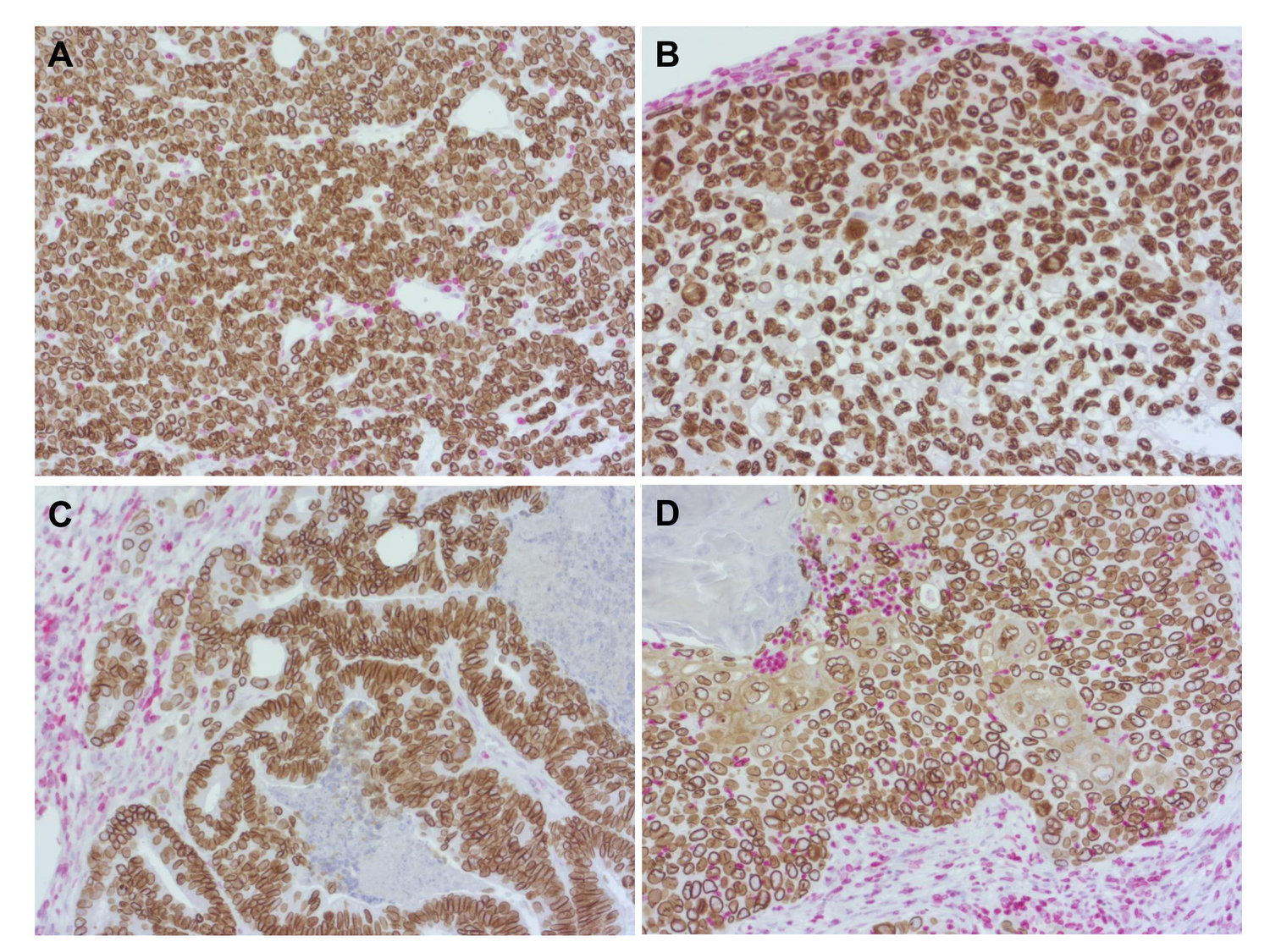
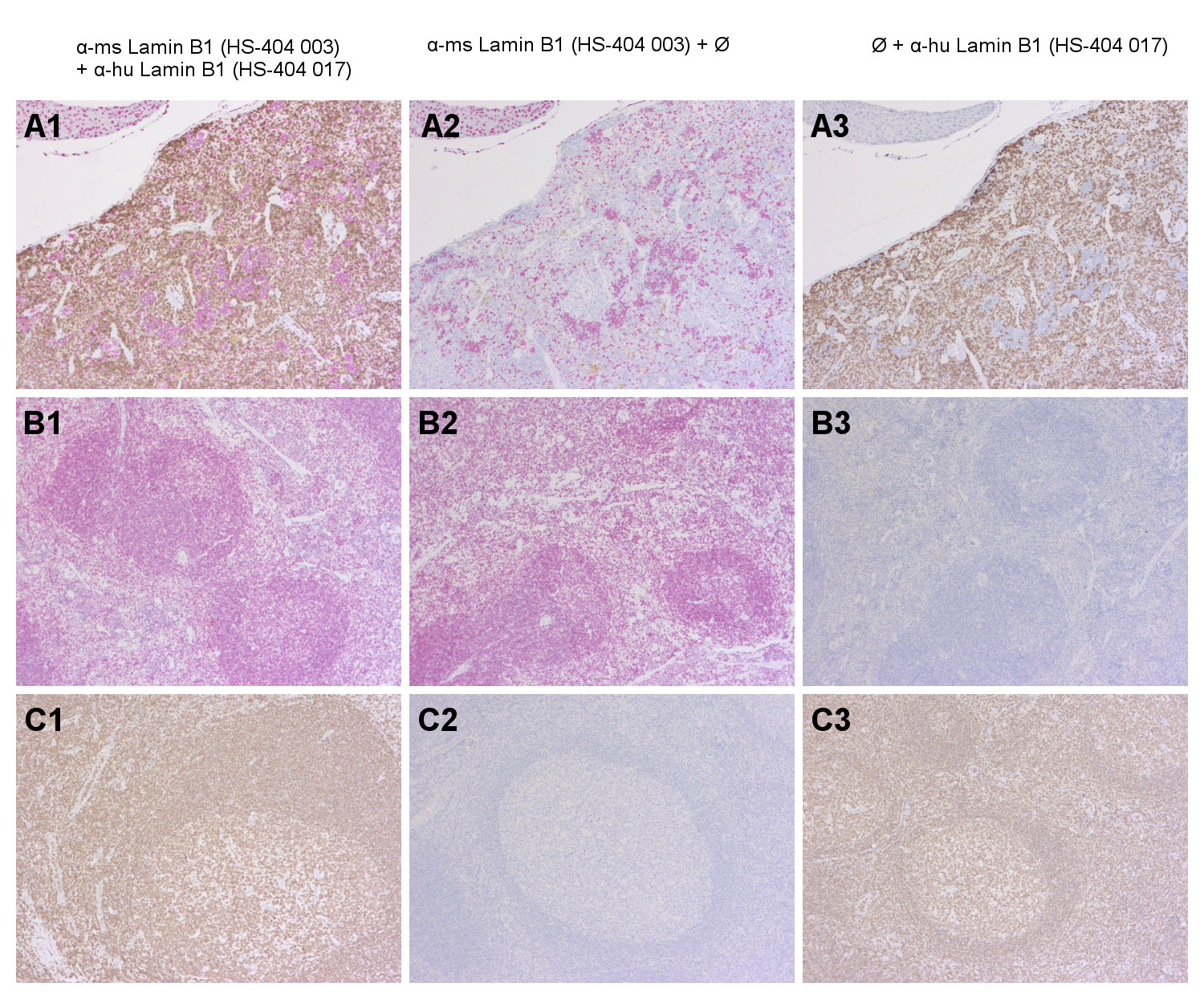
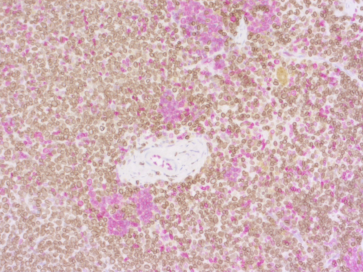
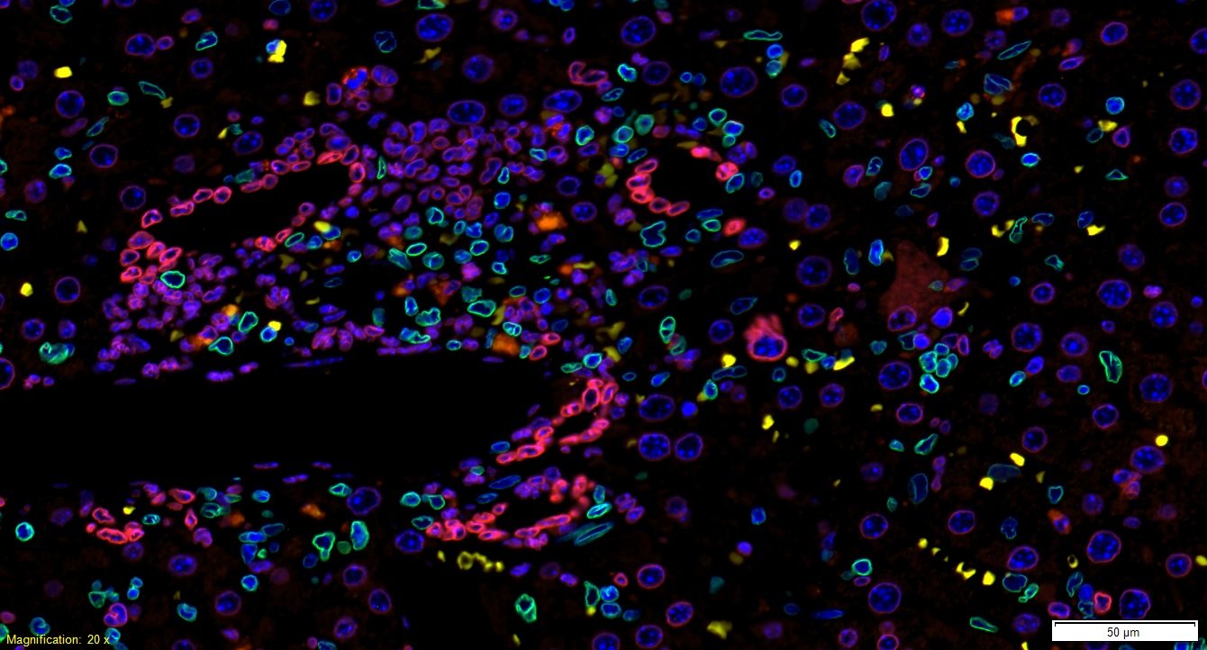
Detection of nuclear Lamin B1 in human FFPE placenta section
Lamin B1 (LMNB1) is an intermediate filament-type protein of the nuclear lamina and is ubiquitously expressed throughout development. It plays important roles in many cellular processes like the distribution of heterochromatin and the regulation of gene expression and splicing. The maintenance of LMNB1 protein levels is required for DNA replication and repair and thus mutations in B-type lamins are usually lethal.
Duplication of the LMNB1 gene causes adult-onset autosomal-dominant leukodystrophy (ADLD), a rare neurological disorder in which overexpression of LMNB1 causes progressive central nervous system demyelination. Improper Lamin B1 expression is often present in tumor cells and decreased levels are observed for example in colon cancer, breast cancer and B-cell malignancies. Lamin B1 loss is also a senescence-associated biomarker and distinguishes senescent from proliferating cells in pre-neoplastic lesions or marks senescent cells in various age-related pathologies.
Certificates
ISO 9001 2015 Quality Management System and Green Lab Platinum certification level for sustaining laboratory processes.


Newsletter
Sign up for our newsletter and get the latest updates and news.