Cat. No.: 525 005
Amount: 50 µg
Price:
$465.00
|
|

|
|
| Cat. No. 525 005 |
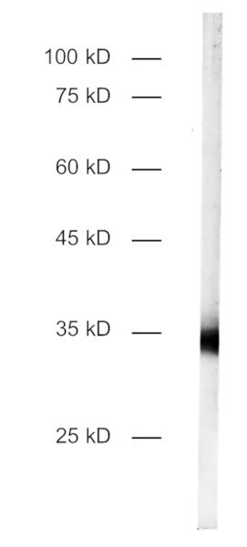
50 µg specific antibody, lyophilized. Affinity purified with the immunogen. Albumin and azide were added for stabilization. For reconstitution add 50 µl H2O to get a 1mg/ml solution in PBS. Then aliquot and store at -20°C to -80°C until use. Antibodies should be stored at +4°C when still lyophilized. Do not freeze! |
| Applications | |
| Immunogen | Synthetic peptide corresponding to residues near the amino terminus of mouse MLC1 (UniProt Id: Q8VHK5) |
| Reactivity |
Reacts with: mouse (Q8VHK5 ), rat (D4ABB2), human (Q15049). Other species not tested yet. |
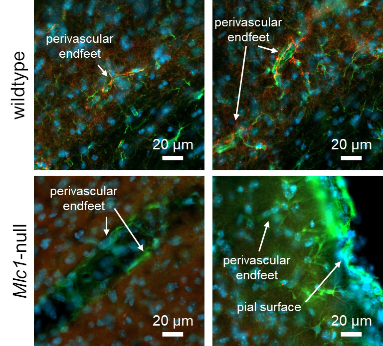
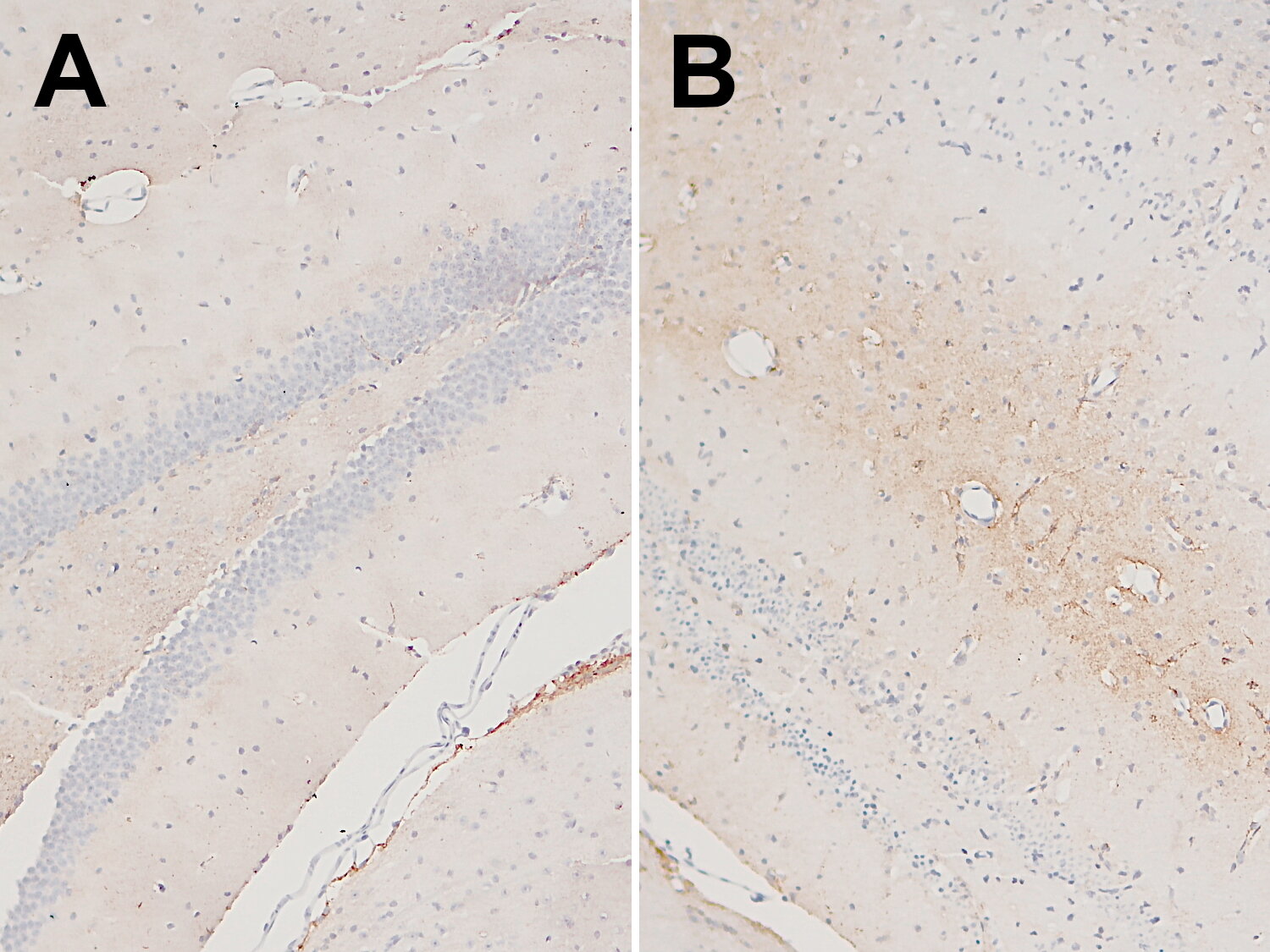
| Specificity | K.O. validated |
| Remarks |
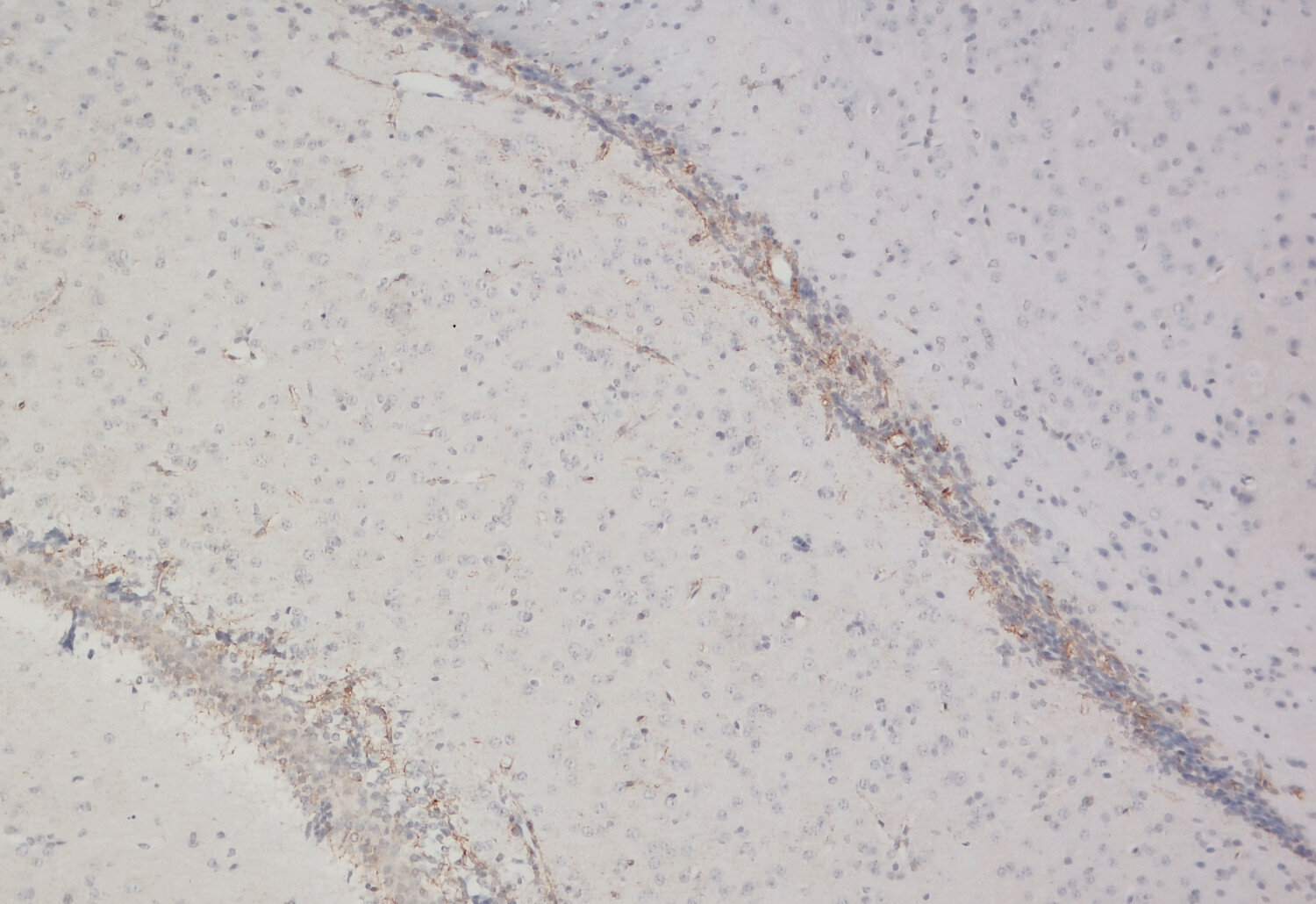
IHC: Antigen retrieval with citrate buffer pH 6 is required. |
| Data sheet | Datasheet 525_005 |
|
|
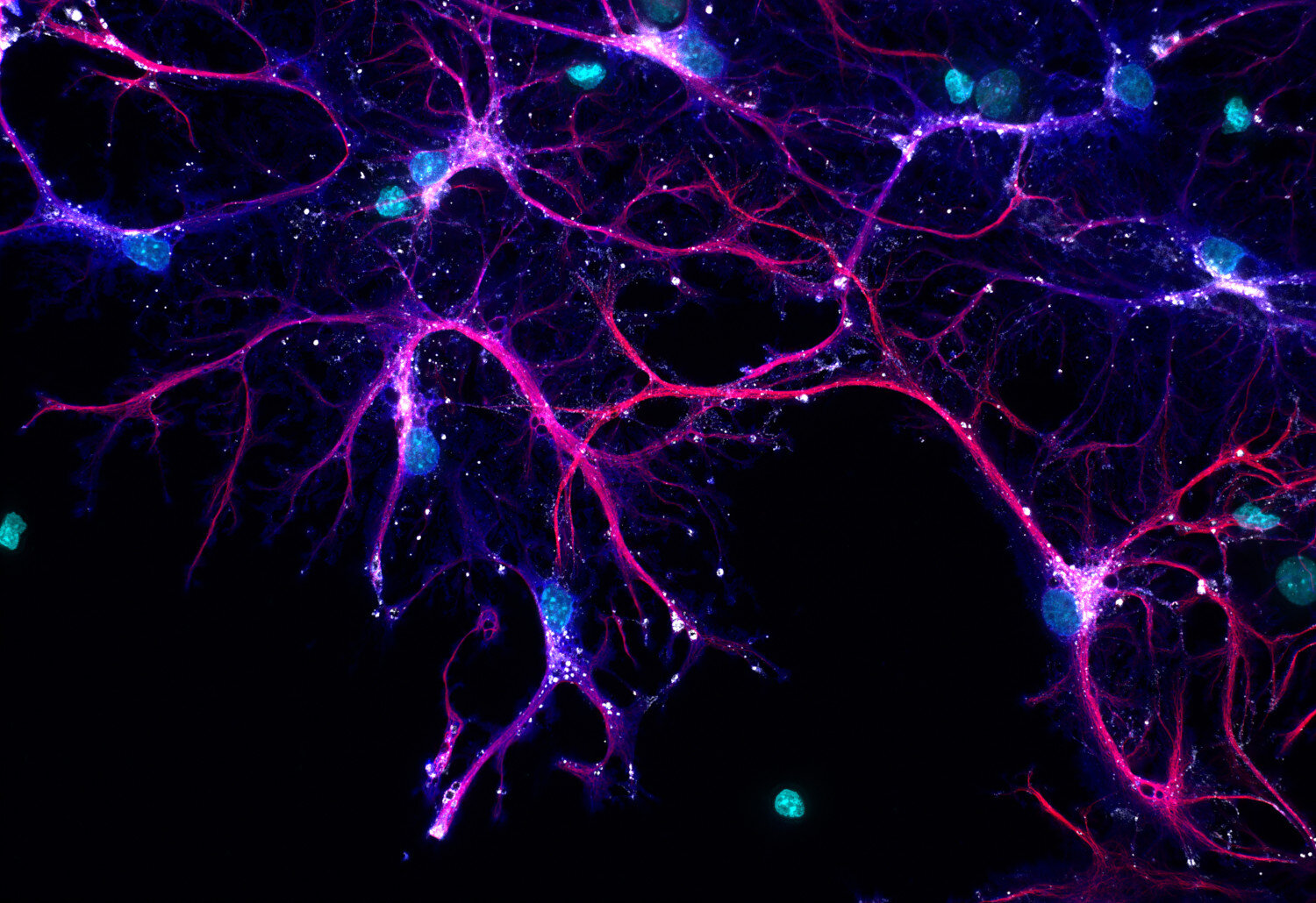
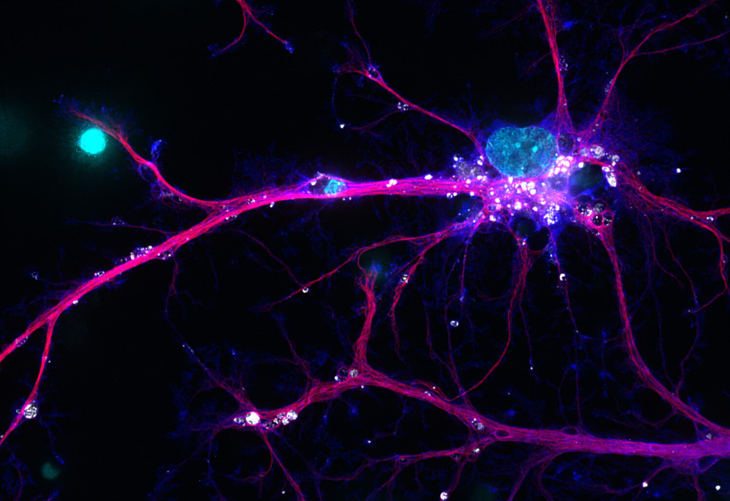
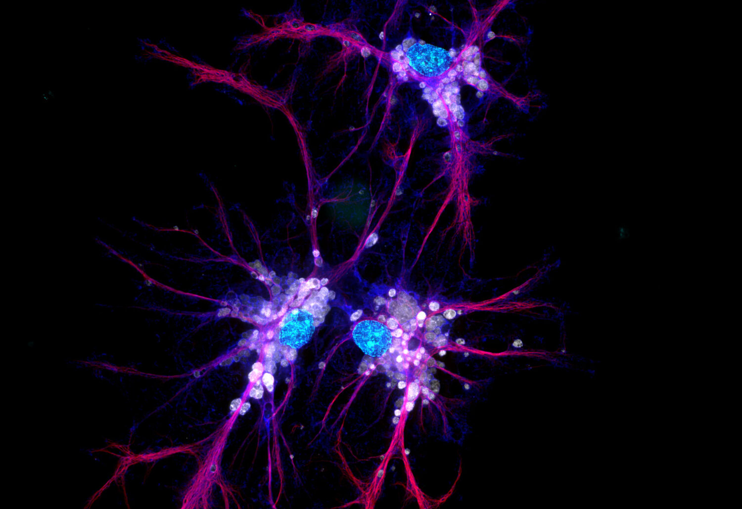
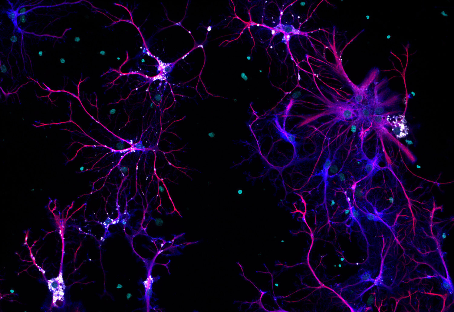
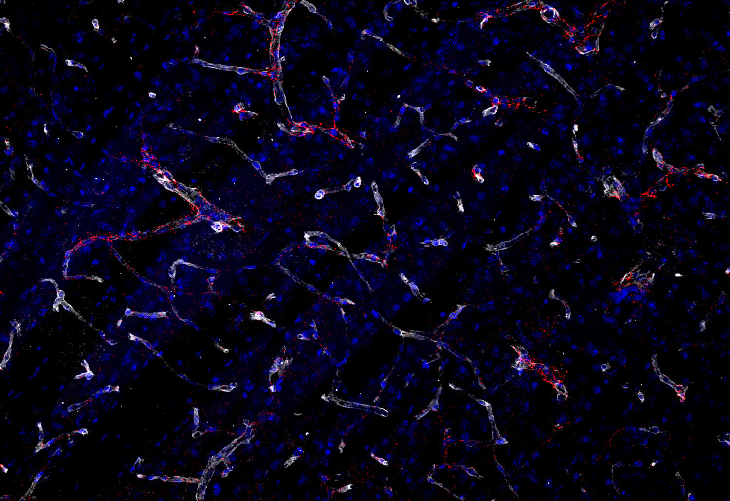
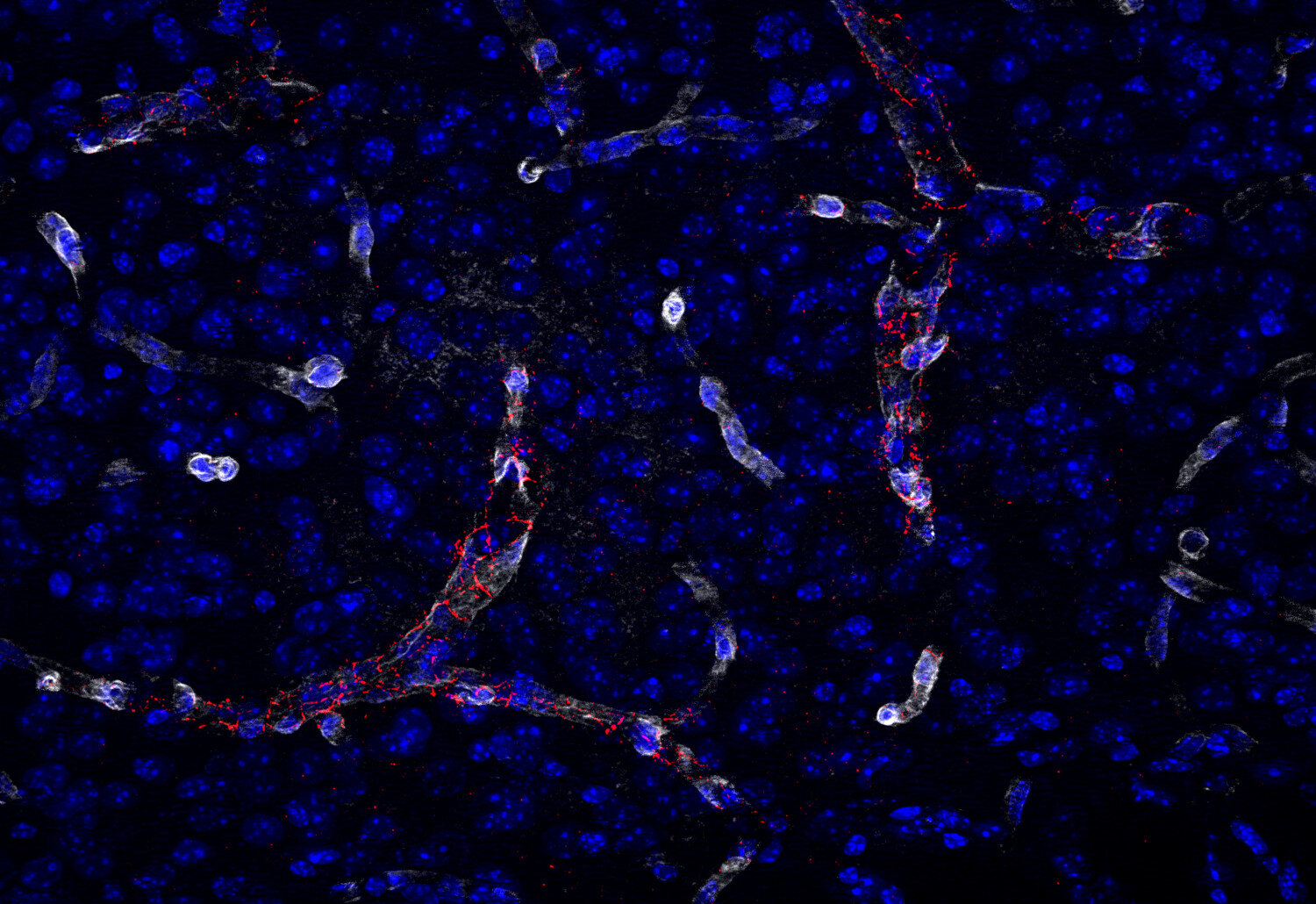
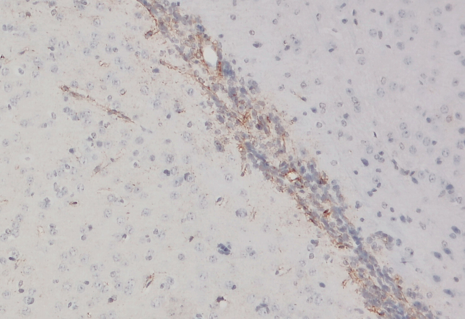
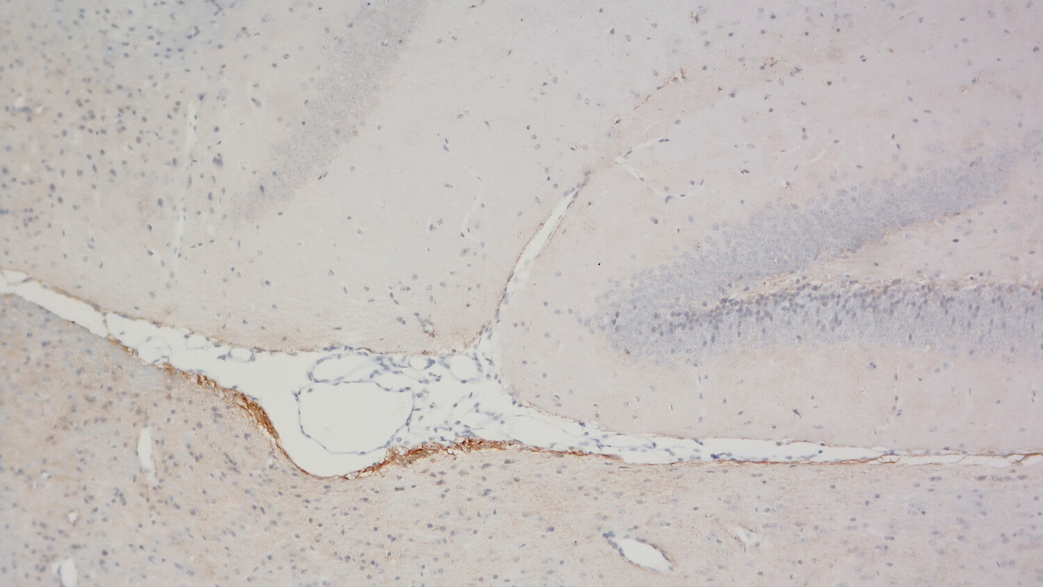
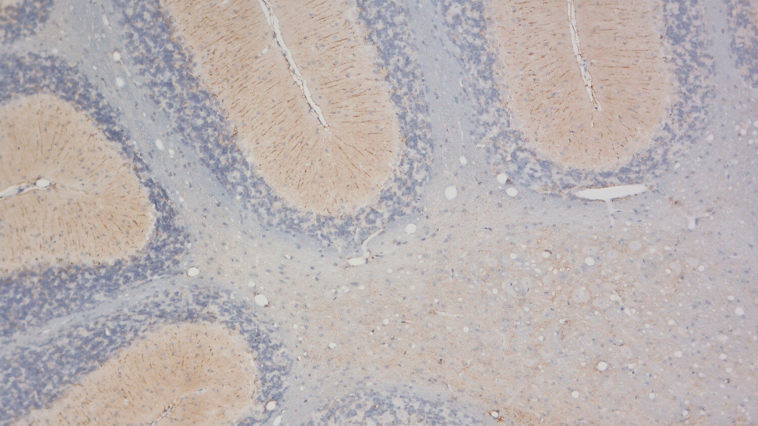
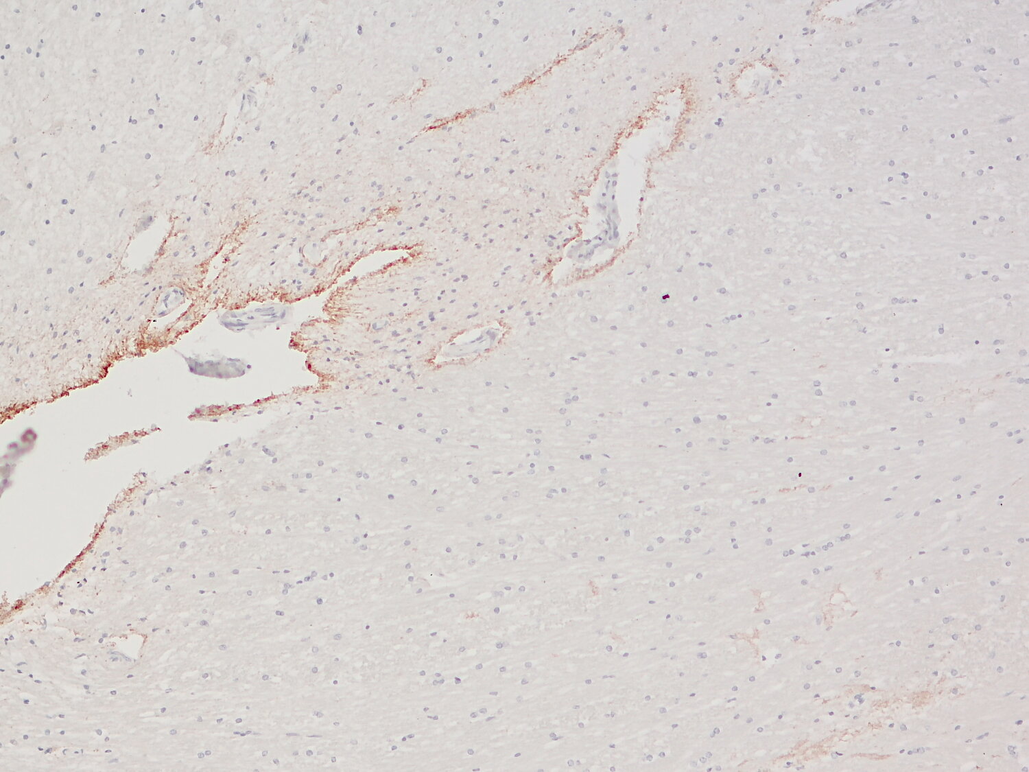
MLC1 is a membrane protein highly expressed in the distal processes of astrocytes, particularly in perivascular, subpial, and subependymal regions. Recessive mutations in MLC1 genes cause Megalencephalic leukoencephalopathy with subcortical cysts (MLC). Additionally, MLC1 upregulation has been observed in brain tissues affected by multiple sclerosis, Alzheimer’s disease, and Creutzfeldt-Jakob disease.
Although its precise function remains unclear, MLC1 interacts with various proteins, including adhesion proteins such as GlialCAM, chloride channels like CLCN2, and gap junction proteins such as connexin 43. It also modulates receptor tyrosine kinases, including EGFR and Axl, by suppressing their enzymatic activity.
MLC1 plays a crucial role in restoring astrocyte homeostasis after inflammation by inhibiting IL-1β–induced inflammatory signaling pathways (pERK, pNF-kB) (3). Experimental in vitro and ex vivo studies suggest that MLC1 contributes to cell volume regulation in astrocytes during osmotic stress by modulating anion channels (VRAC) and Ca²⁺ influx.
Recent findings indicate that loss of MLC1 disrupts astrocyte–excitatory synapse interactions, potentially affecting extracellular glutamate dynamics under conditions of impaired glutamate transporter activity (5). Furthermore, MLC1 stabilizes membrane structure fluctuations in primary astrocytes by regulating actin filament branching (2).
Certificates
ISO 9001 2015 Quality Management System and Green Lab Platinum certification level for sustaining laboratory processes.


Newsletter
Sign up for our newsletter and get the latest updates and news.