Collagen type IV antibody - 462 004

Collagen type IV is a basement membrane protein
Guinea pig polyclonal antiserum
Cat. No.: 462 004
Amount: 100 µl
Price: $370.00
Cat. No. 462 004 100 µl antiserum, lyophilized. For reconstitution add 100 µl H2O, then aliquot and store at -20°C until use.
Antibodies should be stored at +4°C when still lyophilized. Do not freeze!
Applications
 
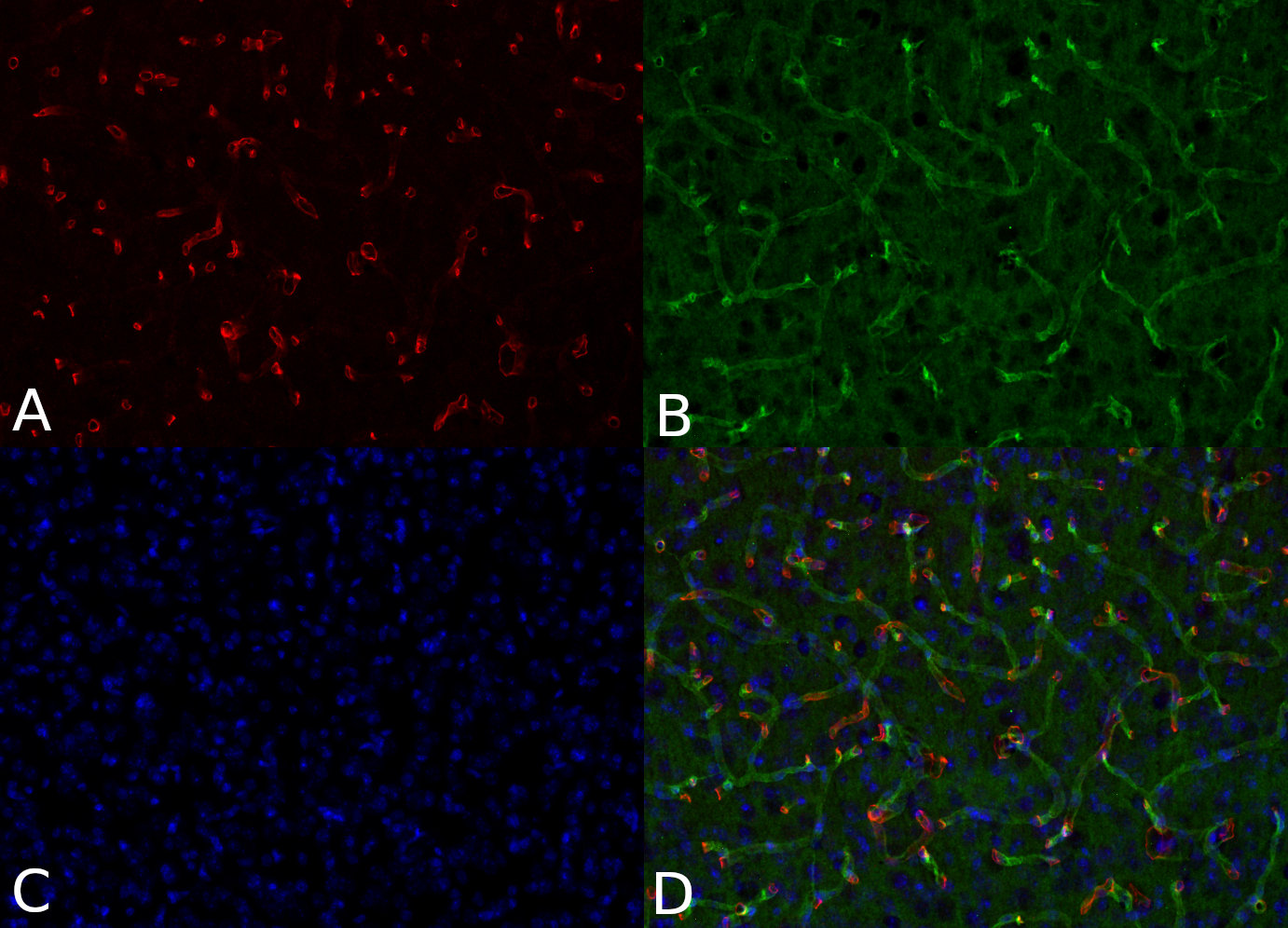
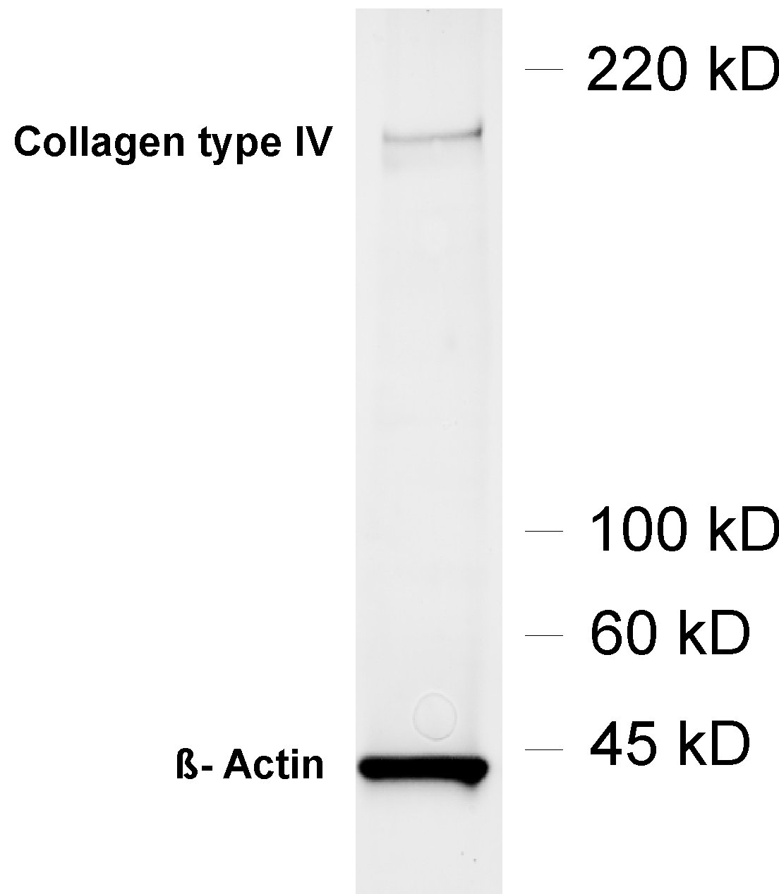
WB: 1 : 1000 (AP staining) gallery
IP: yes
ICC: not tested yet
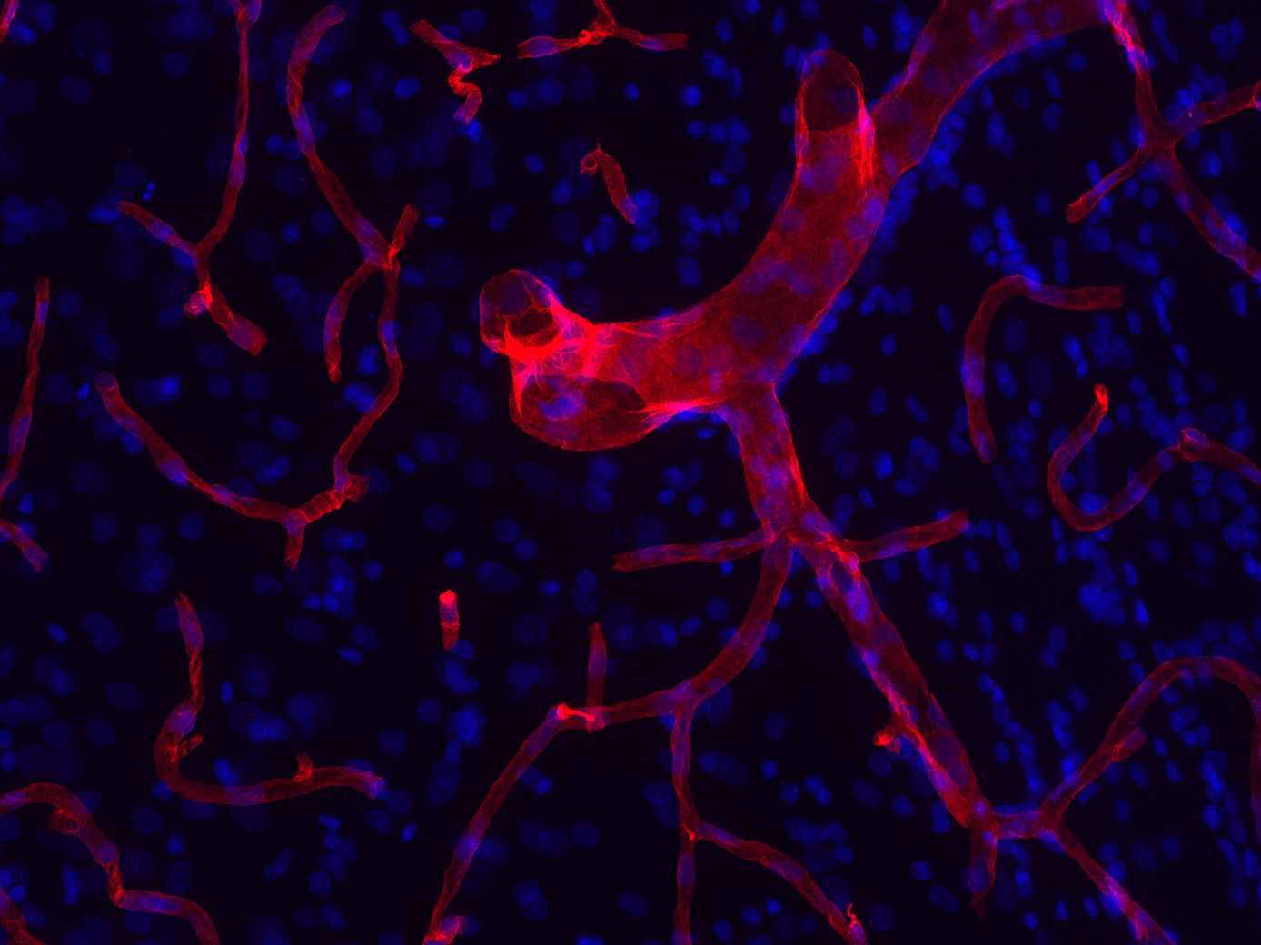
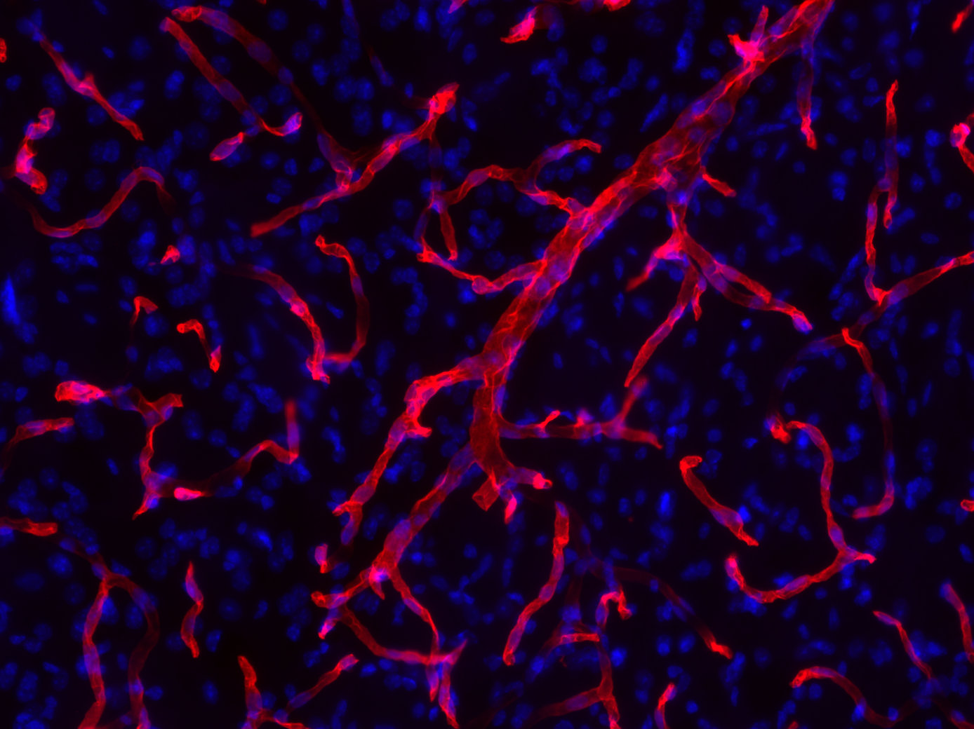
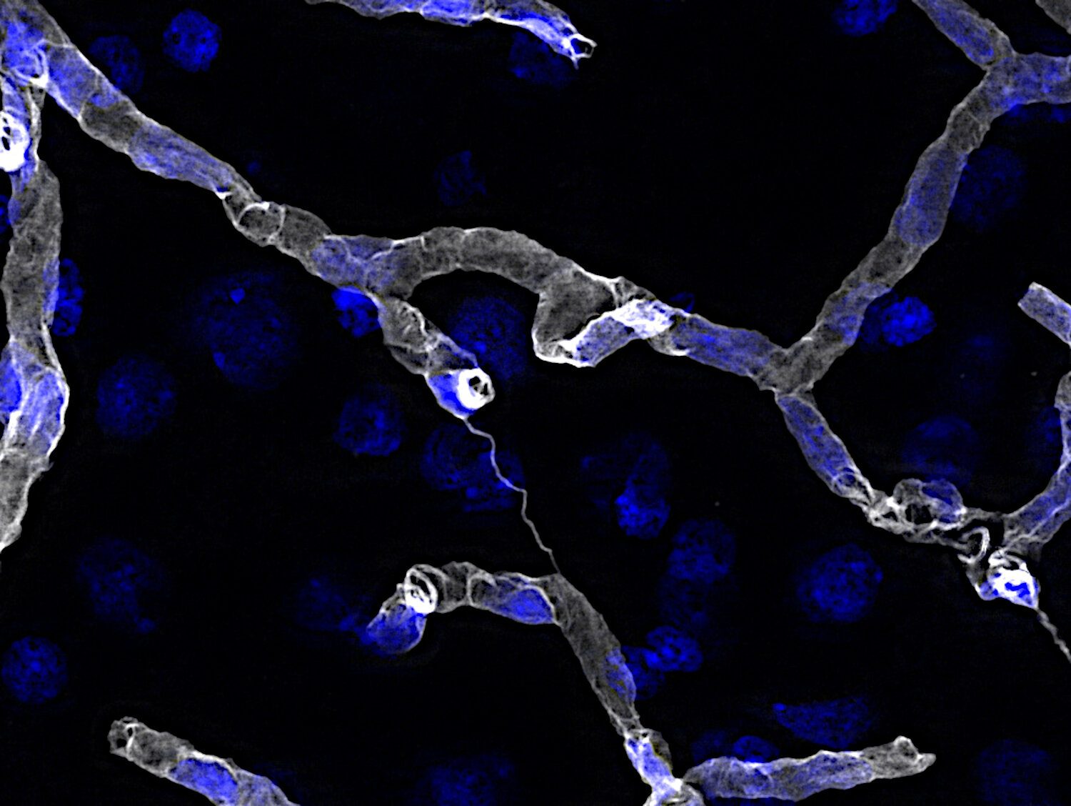
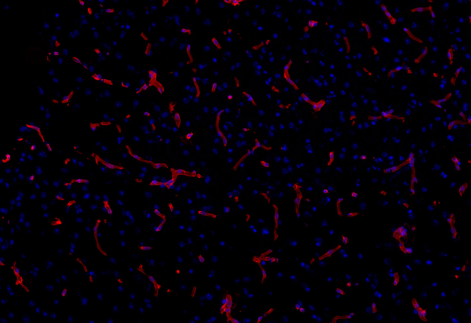
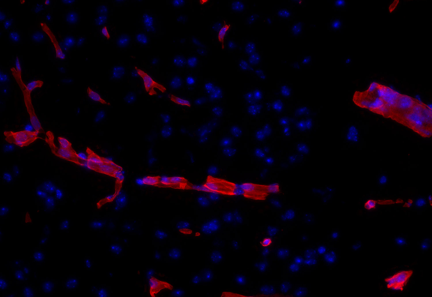
IHC: 1 : 250 up to 1 : 1000 (see remarks) gallery
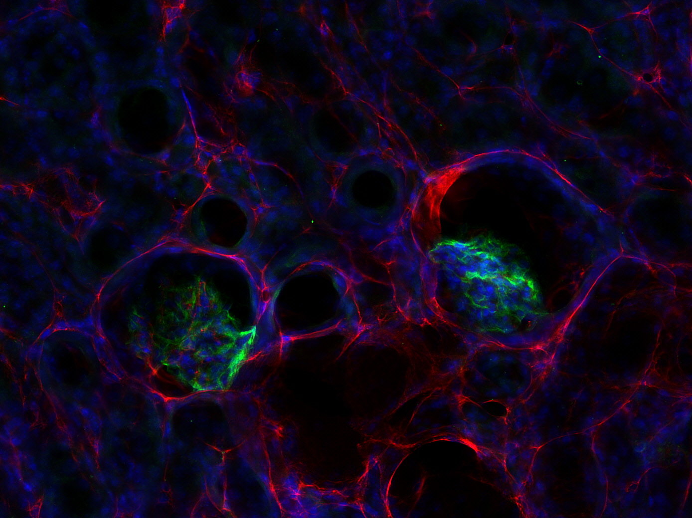
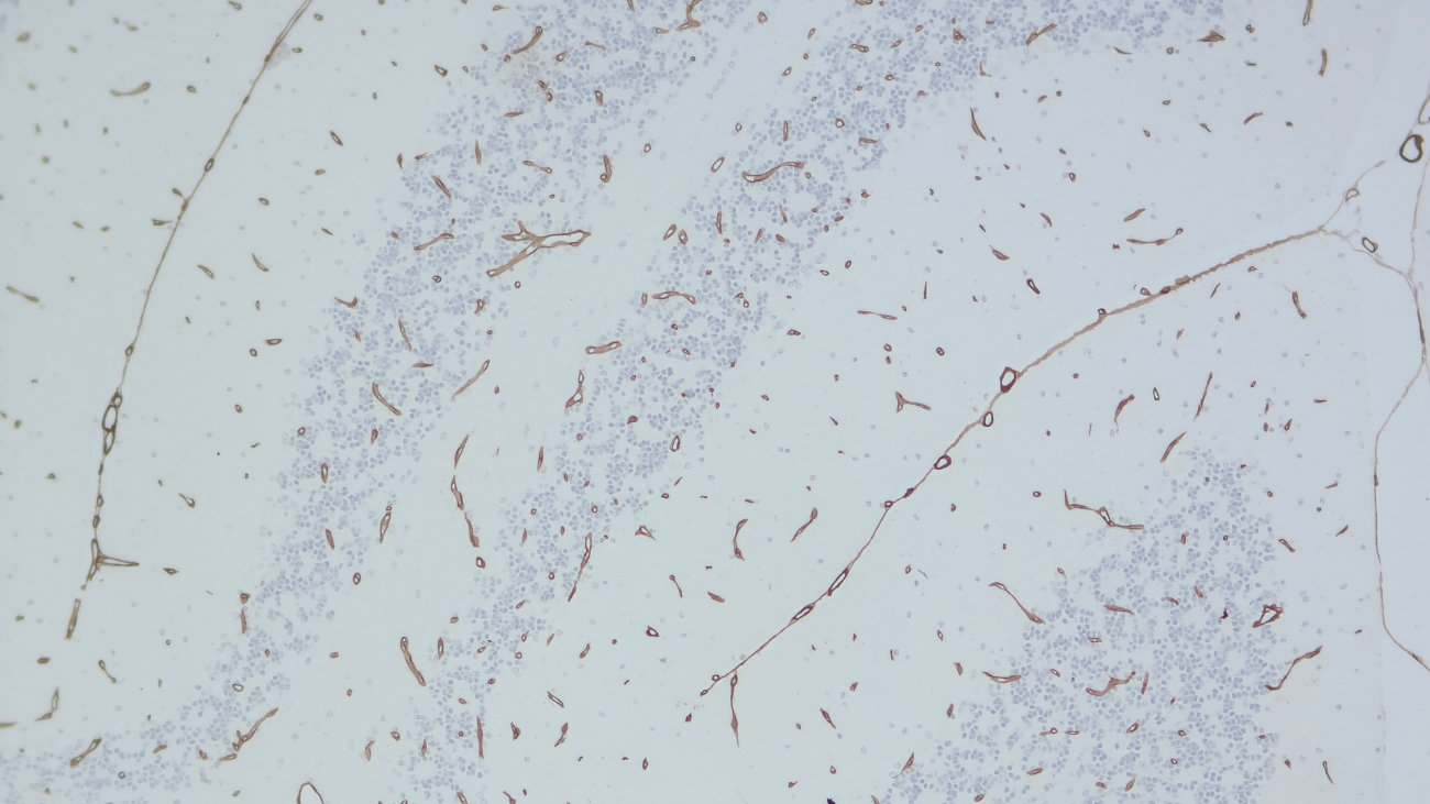
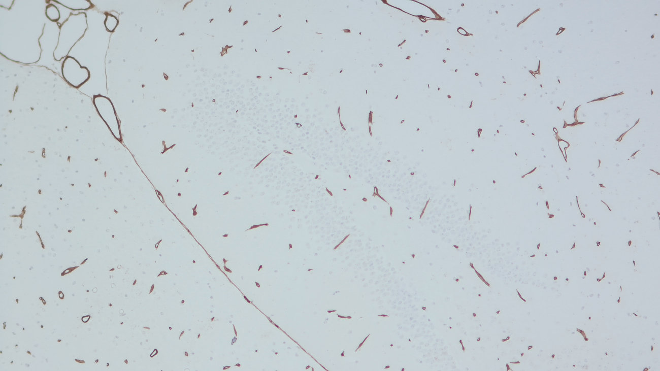
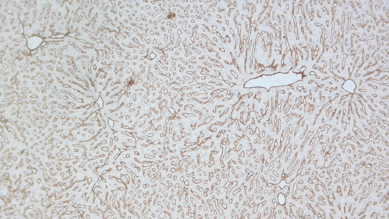
IHC-P (FFPE): 1 : 500 up to 1 : 100 (see remarks) gallery
IHC-Fr: 1 : 500 up to 1 : 1000 (see remarks) gallery
IHC-G: 1 : 500 up to 1 : 1000 (see remarks) gallery

Western blot (WB); separation of proteins by PAGE and subsequent transfer to a membrane. Detection of target molecules is carried out with antibodies. Some antibodies require special sample preparation steps. For details, please refer to the “Remarks” section.

Immunoprecipitation (IP); Immunoisolation or pulldown of a target molecule using an antibody. For details and product specific hints, please refer to the ”Remarks” section.

Immunocytochemistry (ICC) on 4% PFA fixed cells. Immunoreactivity is usually revealed by fluorescence. Some antibodies require special fixation methods. For details, please refer to the “Remarks” section.

Immunohistochemistry (IHC) on 4% PFA perfusion fixed tissue with 24h PFA post fixation. Immunoreactivity is usually revealed by fluorescence or a chromogenic substrate. Some antibodies require special fixation methods or antigen retrieval steps. For details, please refer to the ”Remarks” section.

Immunohistochemistry (IHC-P) of formalin fixed, paraffin embedded (FFPE) tissue (some antibodies require special antigen retrieval steps, please refer to the ”Remarks” section). Immunoreactivity is usually revealed by fluorescence or a chromogenic substrate.

Immunohistochemistry on fresh frozen (IHC-Fr) cryo-tissue-sections. In contrast to standard PFA perfusion fixed tissues, fresh frozen cryo-tissue-sections can be variably postfixed with alcohols, acetone or PFA. Alcohol or acetone fixation is e.g. of advantage for antigens masked by PFA crosslinking. For recommended postfixation, please refer to the ”Remarks” section. Immunoreactivity is usually revealed by fluorescence or a chromogenic substrate.

Immunohistochemistry on glyoxal fixed (IHC-G) tissue. The tissue is perfused with saline and afterwards immersion fixed with a glyoxal solution. For details of the glyoxal solution, please refer to the remarks section. Immunoreactivity is usually revealed by fluorescence.

Immunogen human heterotrimeric Collagen type IV containing two α 1-like (UniProt Id: P02462) and one α 2-like (UniProt Id: P08572) chains.
Reactivity Reacts with: mouse (P02463), rat (F1MA59), human (P02462), human (P08572), mouse (P08122), rat (F1M6Q3).
Other species not tested yet.
Remarks

IHC: Antigen retrieval with pepsin is required.
IHC-P (FFPE): Antigen retrieval with Tris-EDTA buffer pH 9, followed by pepsin treatment, is required for chromogenic detection.
IHC-Fr: 4% formaldehyde/PFA fixation is recommended.
IHC-G: The following fixatives are possible: 3% glyoxal, 9% glyoxal.

Data sheet Data Sheet 462_004

References for Collagen type IV - 462 004

Spatial proteomic analysis in human Alzheimer's disease brains enables identification of microenvironment-dependent microglial cell states.
Sanchez-Molina P, Rosmus DD, Brownell D, Meral M, Gohlich C, Pratapa A, Peymanfar Y, Whitley A, Hou Y, Nikulina N, Bogachuk A, et al.
Nature neuroscience (2026) : . 462 004 IHC-P (FFPE); tested species: human
Cat. No.: 462 004
Amount: 100 µl
Price: $370.00
Spatial proteomic analysis in human Alzheimer's disease brains enables identification of microenvironment-dependent microglial cell states.
Sanchez-Molina P, Rosmus DD, Brownell D, Meral M, Gohlich C, Pratapa A, Peymanfar Y, Whitley A, Hou Y, Nikulina N, Bogachuk A, et al.
Nature neuroscience (2026) : . 462 004 IHC-P (FFPE); tested species: human
Background

Collagen type IV is the most abundant structural component of the basement membrane, a specialized extracellular matrix, that supports epithelial and endothelial cells and separates tissues from their surrounding environments. Collagen type IV consists of polypeptide chains derived from six different alpha chains that assemble into three different collagen type IV isoforms. Unlike fibrillar collagens, collagen IV forms flexible, sheet-like networks that provide structural stability and interact with other basement membrane components, including laminins, nidogens, and proteoglycans (1,2).
The principal functions of collagen IV are to provide mechanical support, maintain basement membrane integrity, regulate cell adhesion, and contribute to tissue organization and cellular signaling. Through its interactions with cell-surface receptors and other extracellular matrix proteins, collagen IV also influences cellular behavior and tissue homeostasis (1).
Dysregulation of collagen IV is associated with several important diseases. Pathogenic variants can cause e.g. diabetic nephropathy, Alport syndrome, and cerebral small-vessel disease as well as increasing the risk of intracerebral hemorrhage and other neurological manifestations (2,3). 

Certificates

ISO 9001 2015 Quality Management System and Green Lab Platinum certification level for sustaining laboratory processes.

Newsletter

Sign up for our newsletter and get the latest updates and news.
 

Please provide a valid E-Mail-Address
Please provide a valid First name
Please provide a valid Last name