Cat. No.: 310 118
Amount: 50 µg
Price:
$420.00
|
|

|
|
| Cat. No. 310 118 |
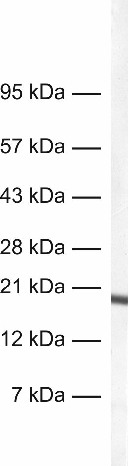
50 µg purified recombinant IgG, lyophilized. Albumin and azide were added for stabilization. For reconstitution add 50 µl H2O to get a 1mg/ml solution in PBS. Then aliquot and store at -20°C to -80°C until use. Antibodies should be stored at +4°C when still lyophilized. Do not freeze! |
| Applications | |
| Clone | Rb330G5 |
| Subtype | IgG1 (κ light chain) |
| Immunogen | Full-length recombinant human MLC-2V protein (UniProt Id: P10916) |
| Epitop |
AA 105 to 111 from human MLC-2V (UniProt Id: P10916) |
| Reactivity |
Reacts with: human (P10916), rat (P08733), mouse (P51667), pig, chicken. Other species not tested yet. |
| Specificity | Specific for MLC-2V, no cross-reactivity to MLC-2A |
| Remarks |
This antibody is a chimeric antibody based on the well known monoclonal mouse antibody clone 330G5. The constant regions of the heavy and light chains have been replaced by rabbit specific sequences. Therefore, the antibody can be used with standard anti-rabbit secondary reagents. The antibody has been expressed in mammalian cells. |
| Data sheet | Datasheet 310_118 |
|
|

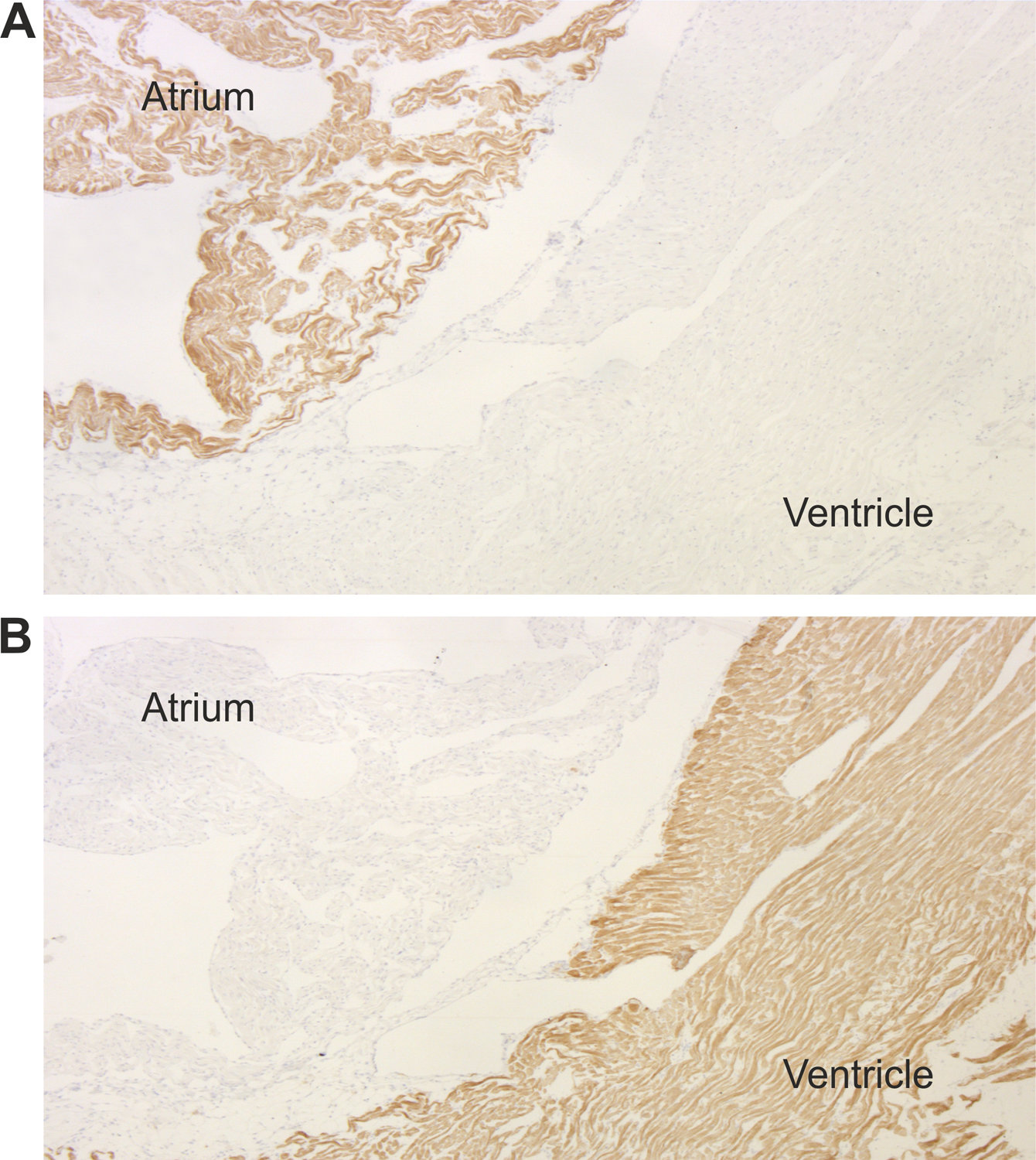
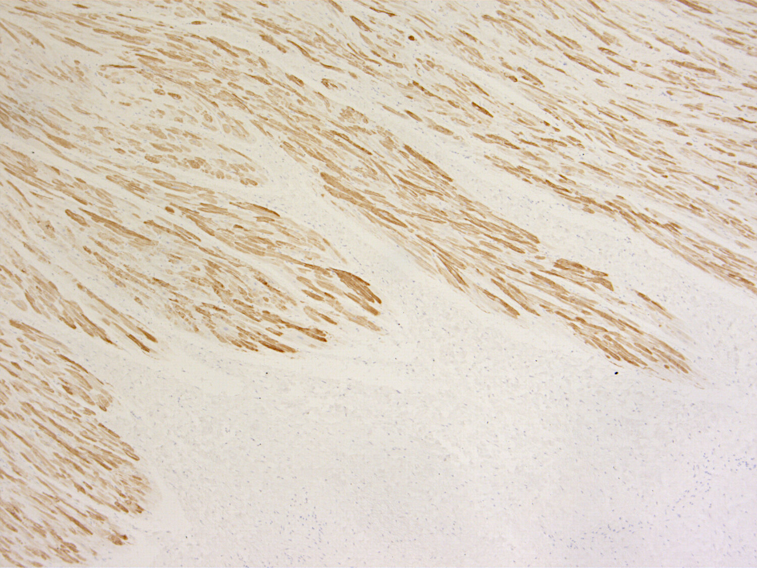
During cardiogenesis two major isoforms of myosin light chain 2 are co-expressed in a tightly regulated manner. MLC-2V is only present in the ventricle while MLC-2A is exclusively expressed in the atrium. Knock out studies revealed that the 2A isoform cannot substitute for the 2V variant in the ventricular chamber.
Recently it has been demonstrated that embryonic and adult stem cells can be differentiated into cardiomyocytes which may generate suitable replacements for damaged heart tissue in the future.
These antibodies are useful tools to distinguish between ventricle and atrium specific cardiomyocytes.
Certificates
ISO 9001 2015 Quality Management System and Green Lab Platinum certification level for sustaining laboratory processes.


Newsletter
Sign up for our newsletter and get the latest updates and news.