Tailor-made Antibodies
and Tools for Life Science
Home | | | | | Technical Support

Rim1/2 antibody - 140 208 K.O.

RIMs are Rab3 effector proteins of the cytomatrix at the active zone
Rabbit monoclonal recombinant IgG
Cat. No.: 140 208
Amount: 50 µg
Price: $420.00
Cat. No. 140 208 
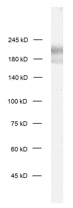
50 µg purified recombinant IgG, lyophilized. Albumin and azide were added for stabilization. For reconstitution add 50 µl H2O to get a 1mg/ml solution in PBS. Then aliquot and store at -20°C to -80°C until use.
Antibodies should be stored at +4°C when still lyophilized. Do not freeze!
Applications
 
WB: 1 : 1000 (AP staining) gallery  
IP: not tested yet
ICC: 1 : 500 gallery  
IHC: 1 : 500 (see remarks) gallery  
IHC-P (FFPE): not recommended
EM: external data (see remarks)

Western blot (WB); separation of proteins by PAGE and subsequent transfer to a membrane. Detection of target molecules is carried out with antibodies. Some antibodies require special sample preparation steps. For details, please refer to the “Remarks” section.

Immunoprecipitation (IP); Immunoisolation or pulldown of a target molecule using an antibody. For details and product specific hints, please refer to the ”Remarks” section.

Immunocytochemistry (ICC) on 4% PFA fixed cells. Immunoreactivity is usually revealed by fluorescence. Some antibodies require special fixation methods. For details, please refer to the “Remarks” section.

Immunohistochemistry (IHC) on 4% PFA perfusion fixed tissue with 24h PFA post fixation. Immunoreactivity is usually revealed by fluorescence or a chromogenic substrate. Some antibodies require special fixation methods or antigen retrieval steps. For details, please refer to the ”Remarks” section.

Immunohistochemistry (IHC-P) of formalin fixed, paraffin embedded (FFPE) tissue (some antibodies require special antigen retrieval steps, please refer to the ”Remarks” section). Immunoreactivity is usually revealed by fluorescence or a chromogenic substrate.

Electron microscopy (EM) is a microscopy technique that detects the scatter of electrons through thin tissue sections. In Immuno-EM the antigen is usually revealed by colloidal gold conjugated secondary antibodies linking electron dense structures to antigen-bound primary antibodies.

Clone Rb53E12
Subtype IgG1 (κ light chain)
Immunogen Recombinant protein corresponding to Zn-finger-domain of rat RIM 2. (UniProt Id: Q9JIS1)
Reactivity Reacts with: mouse (Q99NE5, Q9EQZ7), rat (Q9JIS1, Q9JIR4).
Other species not tested yet.
Specificity K.O. validated
Remarks

This antibody is a chimeric antibody based on the monoclonal rat antibody SY-53E12. The constant regions of the heavy and light chains have been replaced with rabbit specific sequences. The antibody can therefore be used with standard anti-rabbit secondary reagents. The antibody has been expressed in mammalian cells.
IHC: Antigen retrieval with citrate buffer pH 6 is required.
EM: This antibody has been successfully applied and published for this method by customers (see application-specific references).

Data sheet Datasheet 140_208

References for Rim1/2 - 140 208

Presynaptic cAMP-PKA-mediated potentiation induces reconfiguration of synaptic vesicle pools and channel-vesicle coupling at hippocampal mossy fiber boutons.
Kim O, Okamoto Y, Kaufmann WA, Brose N, Shigemoto R, Jonas P
PLoS biology (2024) 2211: e3002879. 140 208 EM; tested species: mouse,rat
Cat. No.: 140 208
Amount: 50 µg
Price: $420.00
Presynaptic cAMP-PKA-mediated potentiation induces reconfiguration of synaptic vesicle pools and channel-vesicle coupling at hippocampal mossy fiber boutons.
Kim O, Okamoto Y, Kaufmann WA, Brose N, Shigemoto R, Jonas P
PLoS biology (2024) 2211: e3002879. 140 208 EM; tested species: mouse,rat
Background

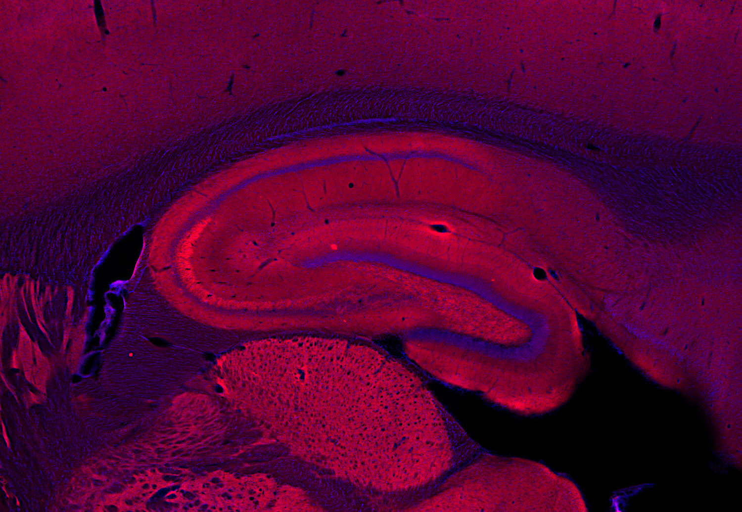
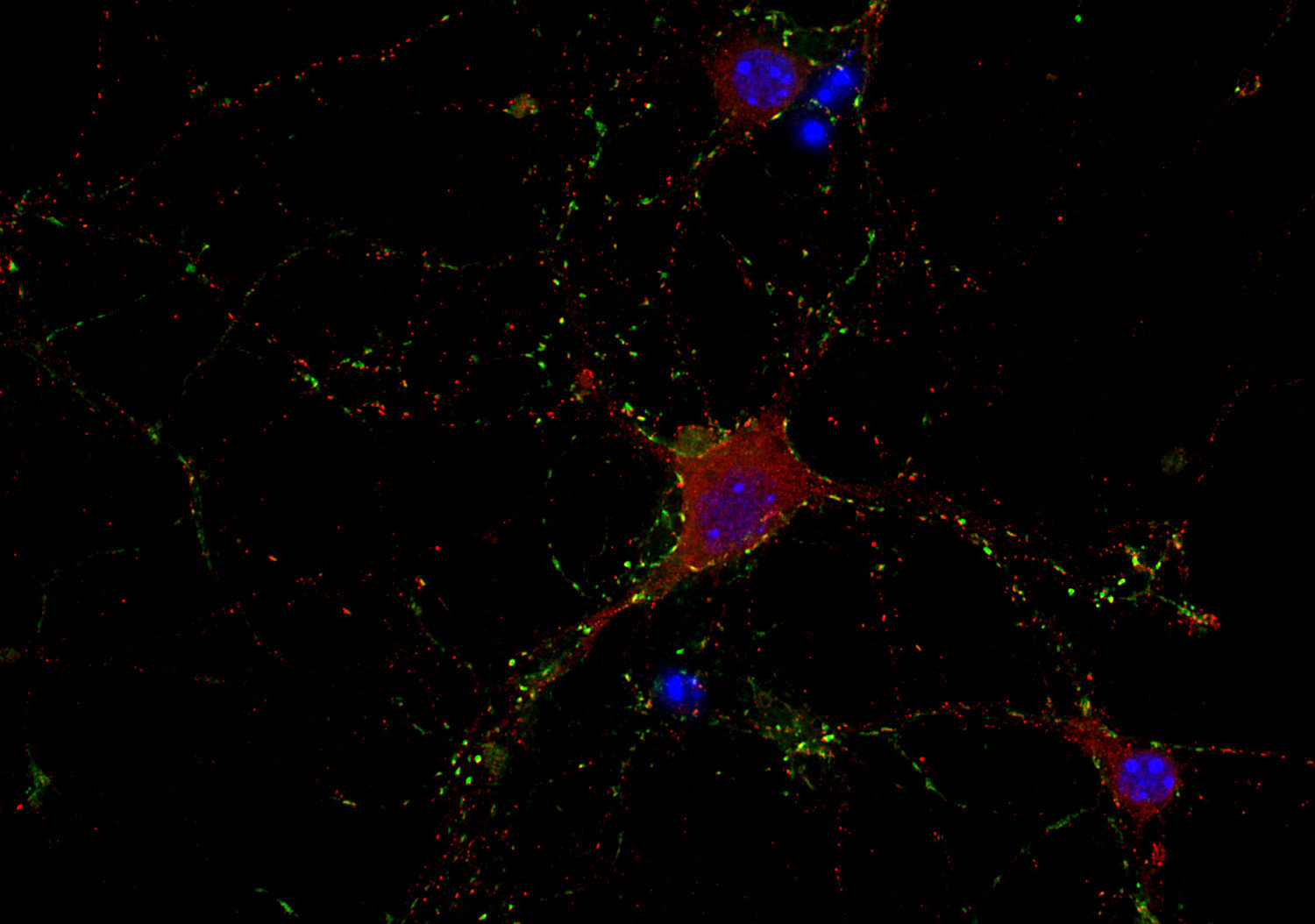
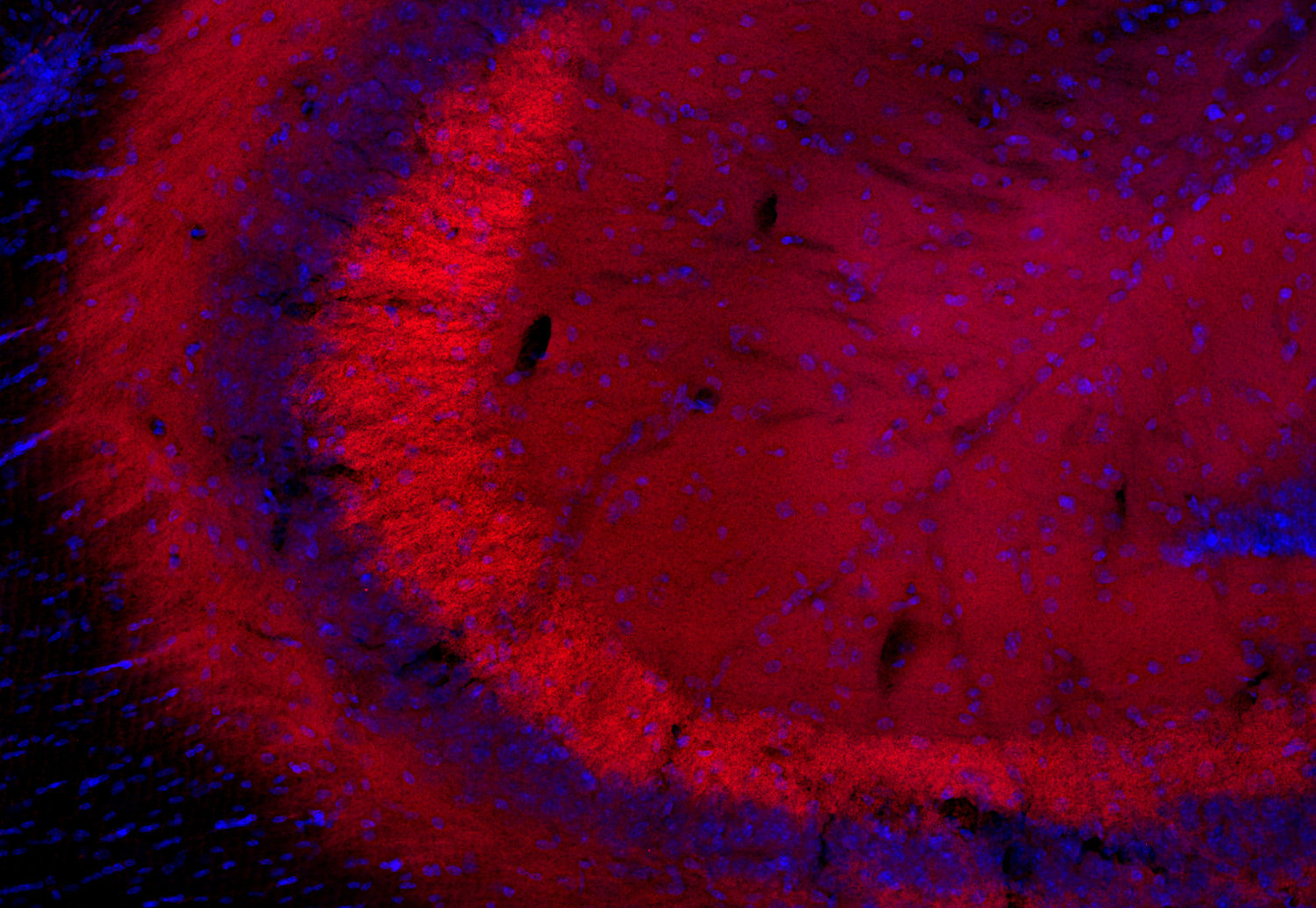
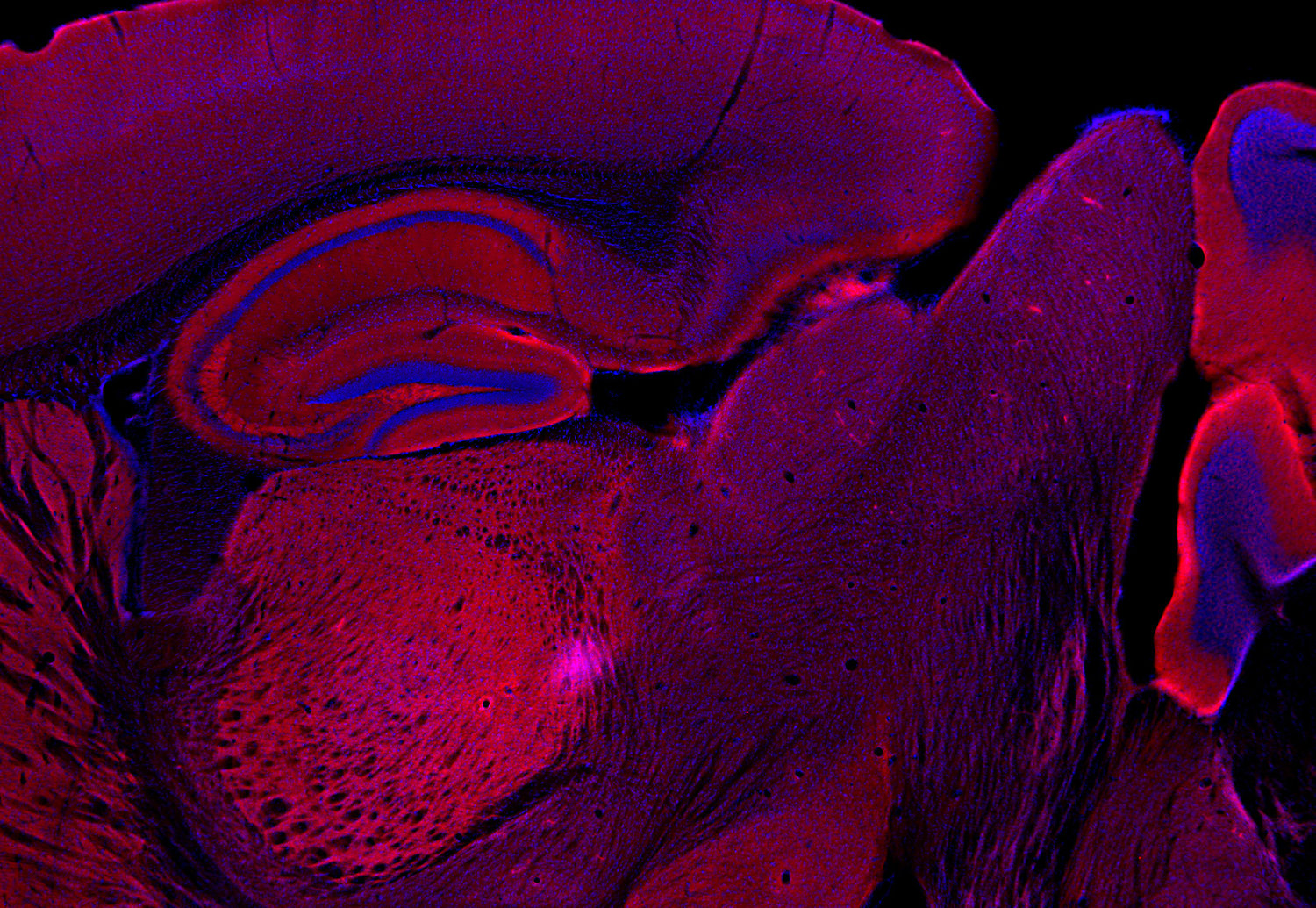
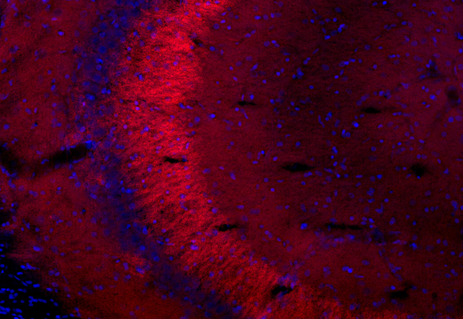
RIMs are presynaptic active zone proteins that regulate Ca2+ triggered release of neurotransmitters. RIM 1α and RIM 2α are composed of an N-terminal zinc-finger domain, a central PDZ domain and two C-terminal C2 domains that are seperated by long alternatively spliced sequences.
RIM 1α is a putative Rab 3a effector and has been shown to interact with other active zone proteins like Munc13-1, ERC 1b, ERC 2 and α-liprins. Deletion of RIM 1α in mice impaired neurotransmitter release without changing the structure of the synapse.
RIM 2β consists of a specific N-terminus, the central PDZ domain and the C-terminal C2 domains. The mRNA for RIM 2β is transcribed from an internal promoter of the RIM 2α gene.
Shorter variants of RIM 2 which comprise only the C-terminal C2B domain and some flanking regions are referred to as NIM 2 / RIM 2γ and NIM 3 / RIM 3γ.

Certificates

ISO 9001 2015 Quality Management System and Green Lab Platinum certification level for sustaining laboratory processes.

Newsletter

Sign up for our newsletter and get the latest updates and news.
 

Please provide a valid E-Mail-Address
Please provide a valid First name
Please provide a valid Last name