Cat. No.: 107 108
Amount: 50 µg
Price:
$420.00
|
|

|
|
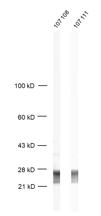
| Cat. No. 107 108 |
50 µg purified recombinant IgG, lyophilized. Albumin and azide were added for stabilization. For reconstitution add 50 µl H2O to get a 1mg/ml solution in PBS. Then aliquot and store at -20°C to -80°C until use. Antibodies should be stored at +4°C when still lyophilized. Do not freeze! |
| Applications | |
| Clone | Rb42.2 |
| Subtype | IgG1 (κ light chain) |
| Immunogen | Full length rat recombinant Rab3a (UniProt Id: P63012) |
| Epitop |
AA 95 to 151 from rat Rab3a (UniProt Id: P63012) |
| Reactivity |
Reacts with: mouse (P63011), rat (P63012), human (P20336). Other species not tested yet. |
| Specificity | K.O. validated |
| Remarks |
This antibody is a chimeric antibody based on the well known monoclonal mouse antibody clone 42.2. The constant regions of the heavy and light chains have been replaced by rabbit specific sequences. Therefore, the antibody can be used with standard anti-rabbit secondary reagents. The antibody has been expressed in mammalian cells. |
| Data sheet | Datasheet 107_108 |
|
|
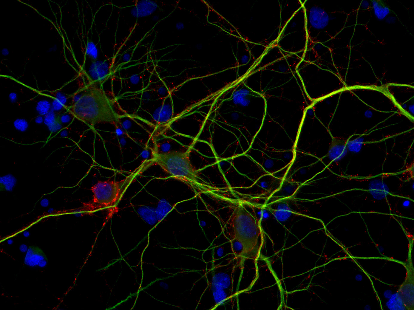
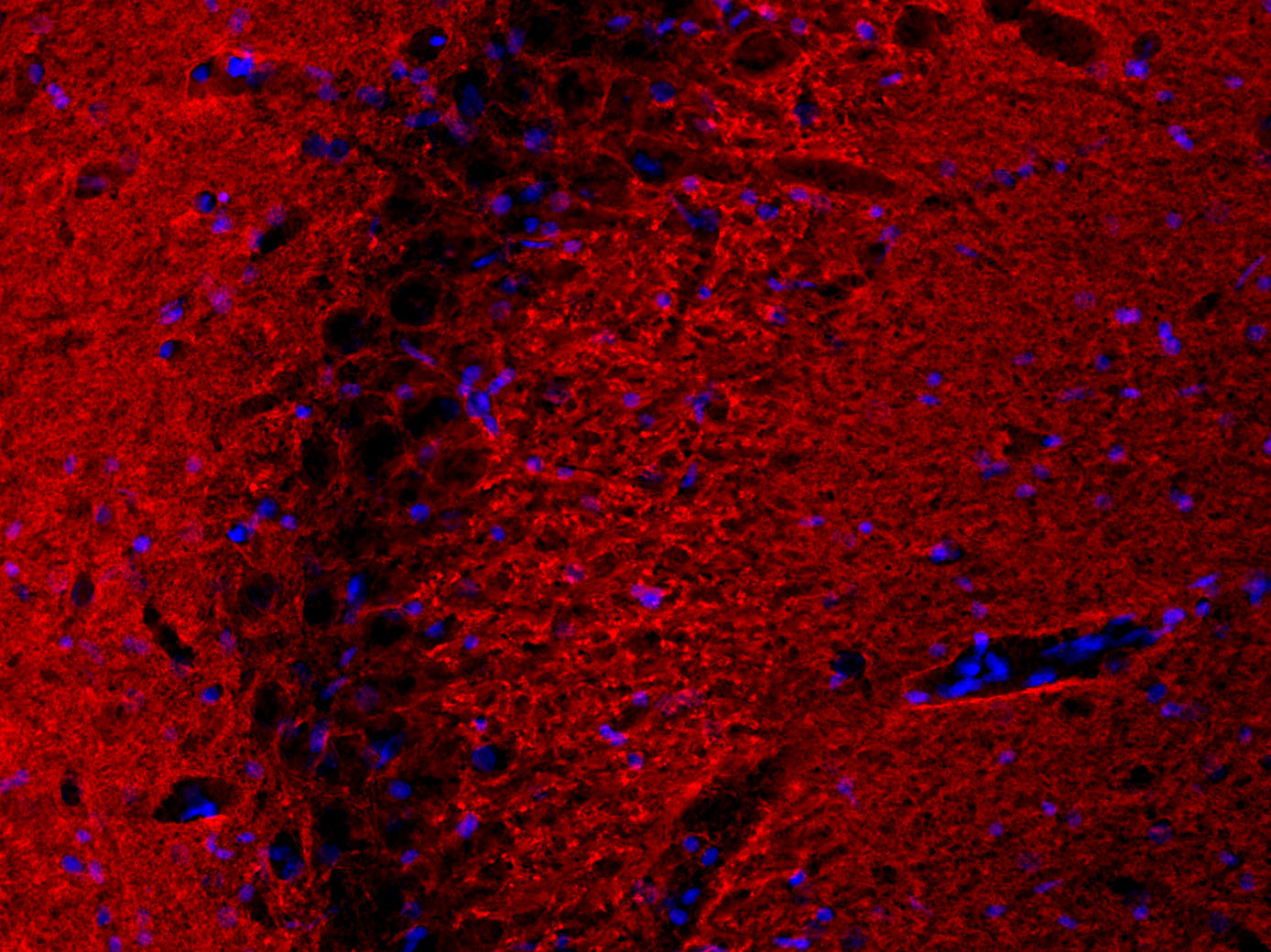
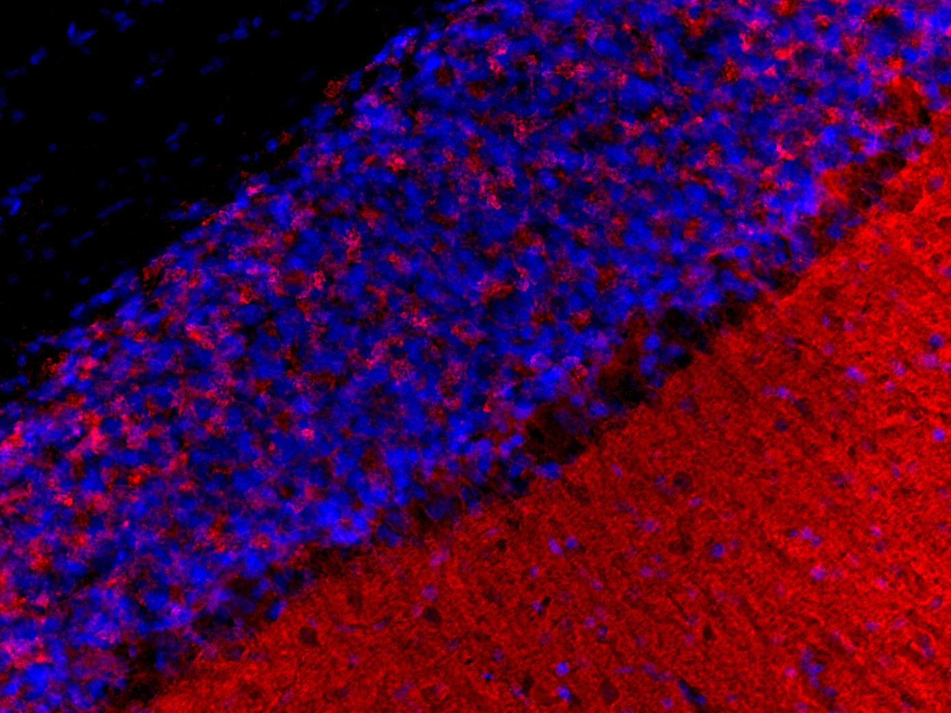
Rab3 proteins belong to the Rab family, a subset of the Ras-related superfamily of small monomeric GTPases. There are four known isoforms: Rab3a, Rab3b, Rab3c, and Rab3d (1, 2). Rab3a and Rab3c are primarily found in neuronal and neuroendocrine cells, whereas Rab3b and Rab3d are predominantly expressed in non-neuronal tissues such as the parotid gland, pancreas, mast cells, and adipose tissue (2, 3).
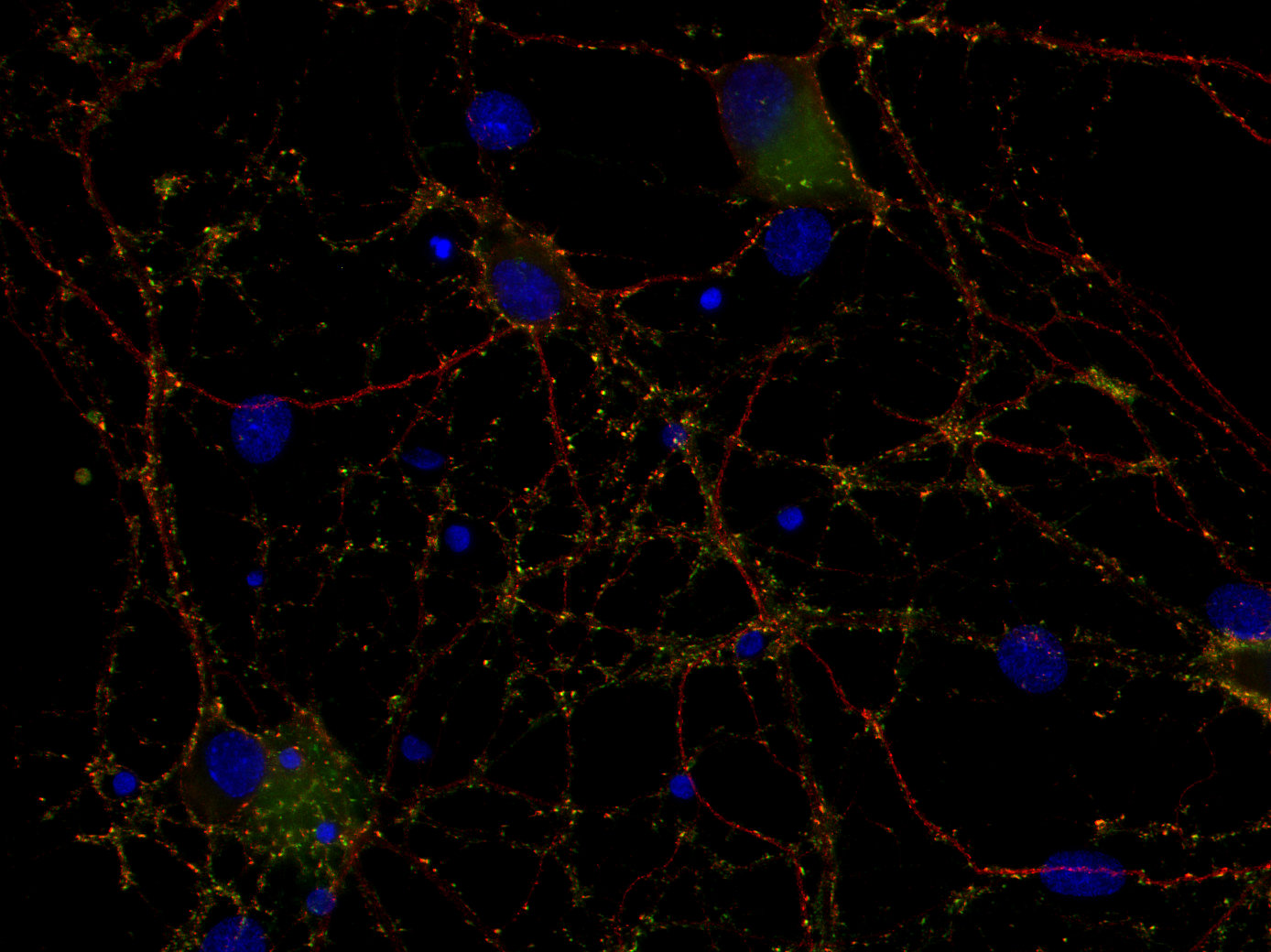
Rab3a, the most abundant and well-characterized isoform, is highly enriched in synaptic vesicles. It regulates vesicle transport, docking, fusion, and Ca²⁺-dependent neurotransmitter release by cycling between a GDP-bound inactive state and a GTP-bound vesicle-associated active state, interacting with other synaptic proteins in the process (1, 2).
Unlike integral membrane proteins of synaptic vesicles, Rab3a and Rab3c are absent from the Golgi complex, preventing immunostaining of the axo-dendritic region, which can occur with proteins such as synaptophysin, synaptobrevin/VAMP, or synaptogyrin (1).
Beyond the nervous system, Rab3a is also expressed in the pancreas, where it is predominantly localized to β-cells of the islets of Langerhans. It plays a crucial role in regulated insulin secretion, while Rab3d is primarily involved in exocrine pancreatic enzyme secretion (3).
Certificates
ISO 9001 2015 Quality Management System and Green Lab Platinum certification level for sustaining laboratory processes.


Newsletter
Sign up for our newsletter and get the latest updates and news.