Cat. No.: 105 311AF
Amount: 100 µg
Price:
$470.00
|
|

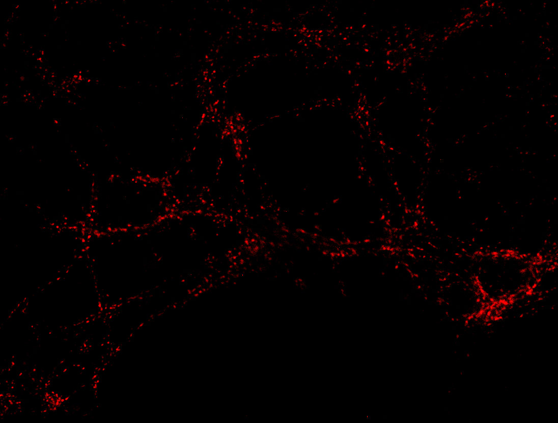
pH-sensitive probe fluorescent only under acidic conditions
|
|
| Cat. No. 105 311AF |
100 µg purified IgG, lyophilized, fluorescence-labeled with
AcidiFluor Orange.
Albumin was added for stabilization. For reconstitution add 100 µl H2O to get a 1mg/ml solution in PBS. Either add 1:1 (v/v) glycerol, then aliquot and store at -20°C until use, or store aliquots at -80°C without additives. Reconstitute immediately upon receipt! Avoid bright light when working with the antibody to minimize photo bleeching of the fluorescent dye. |
| Applications |
IP: N/A ICC: 1 : 100 up to 1 : 500 (see remarks) gallery IHC: not tested yet IHC-P (FFPE): not tested yet EM: N/A ELISA: N/A FACS: not tested yet |
| Label | AcidiFluor Orange |
| Clone | 604.2 |
| Subtype | IgG1 |
| Immunogen | Synthetic peptide corresponding to residues near the amino terminus of rat Synaptotagmin1 (UniProt Id: P21707) |
| Reactivity |
Reacts with: rat (P21707). No signal: mouse (P46096), zebrafish. Other species not tested yet. |
| Remarks |
ICC: This antibody can only be used for labeling of recycling synaptic vesicles in living neurons. It is not recommended for the staining of fixed cells. |
| Data sheet | Datasheet 105_311af |
|
|
Synaptotagmin1, also known as p65, is an integral membrane glycoprotein of neuronal synaptic vesicles and secretory granules of neuroendocrine cells that is widely (but not ubiquitously) expressed in the central and peripheral nervous system. It has a variable N-terminal domain that is exposed to the lumen of the vesicle and a conserved cytoplasmic tail that contains two Ca2+-binding C2-domains.
Ca2+-binding to synaptotagmin triggers exocytosis of synaptic vesicles, thus linking Ca2+-influx during depolarization to neurotransmitter release.
Lumenal antibodies were used in living neurons to label synaptic vesicles from the outside via endocytotic uptake.
For more information on protein expression pattern, please refer to the overview image in our SYSY Antibodies ATLAS.
Certificates
ISO 9001 2015 Quality Management System and Green Lab Platinum certification level for sustaining laboratory processes.


Newsletter
Sign up for our newsletter and get the latest updates and news.