Cat. No.: 399 003
Amount: 50 µg
Price:
$380.00
|
|

|
|
| Cat. No. 399 003 |
50 µg specific antibody, lyophilized. Affinity purified with the immunogen. Albumin was added for stabilization. For reconstitution add 50 µl H2O to get a 1mg/ml solution in PBS. Then aliquot and store at -20°C to -80°C until use. Antibodies should be stored at +4°C when still lyophilized. Do not freeze! |
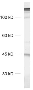
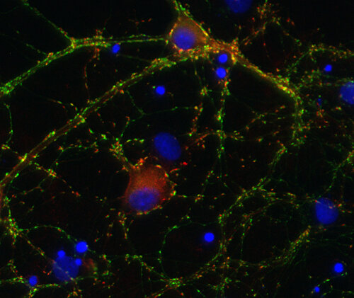
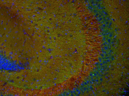
| Applications | |
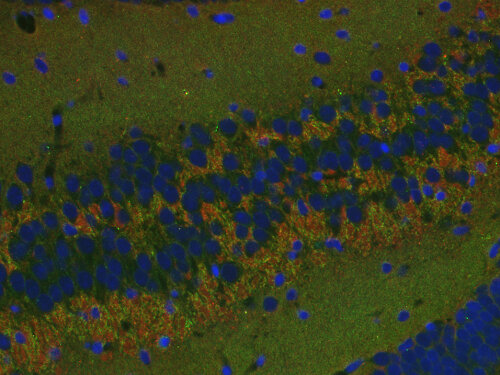
| Immunogen | Synthetic peptide corresponding to AA 290 to 302 from rat Spinophilin (UniProt Id: O35274) |
| Reactivity |
Reacts with: rat (O35274), mouse (Q6R891). Other species not tested yet. |
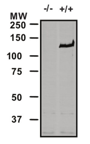
| Specificity | Specific for Spinophilin isoform1. Does not detect isoform 2. K.O. validated |
| Matching control protein/peptide | 399-0P |
| Remarks |
DNA-PAINT: This antibody has been successfully applied and published for this method by customers (see application-specific references). |
| Data sheet | Datasheet 399_003 |
|
|
Certificates
ISO 9001 2015 Quality Management System and Green Lab Platinum certification level for sustaining laboratory processes.


Newsletter
Sign up for our newsletter and get the latest updates and news.