Cat. No.: N1602-At488-L
Amount: 200 µl
Price:
$540.00
|
|

This product was developed by |
Camelid single domain antibodies (sdAbs) consist only of one antigen binding site of an Alpaca heavy chain antibody. With only ~15 kDa, these Tags are about 10-times smaller than conventional IgG antibody molecules.
|
|
| Cat. No. N1602-At488-L |
200 µl purified antibody, lyophilized from PBS, fluorescence-labeled with
ATTO® 488.
Albumin was added for stabilization. For reconstitution refer to the NanoTag reconstitution and storage instructions. Reconstitute immediately upon receipt! Avoid bright light when working with the antibody to minimize photo bleeching of the fluorescent dye. |
| Applications | |
| Label | ATTO 488, two fluorophores coupled to one FluoTag |
| Clone | Nb9 |
| Subtype | single domain |
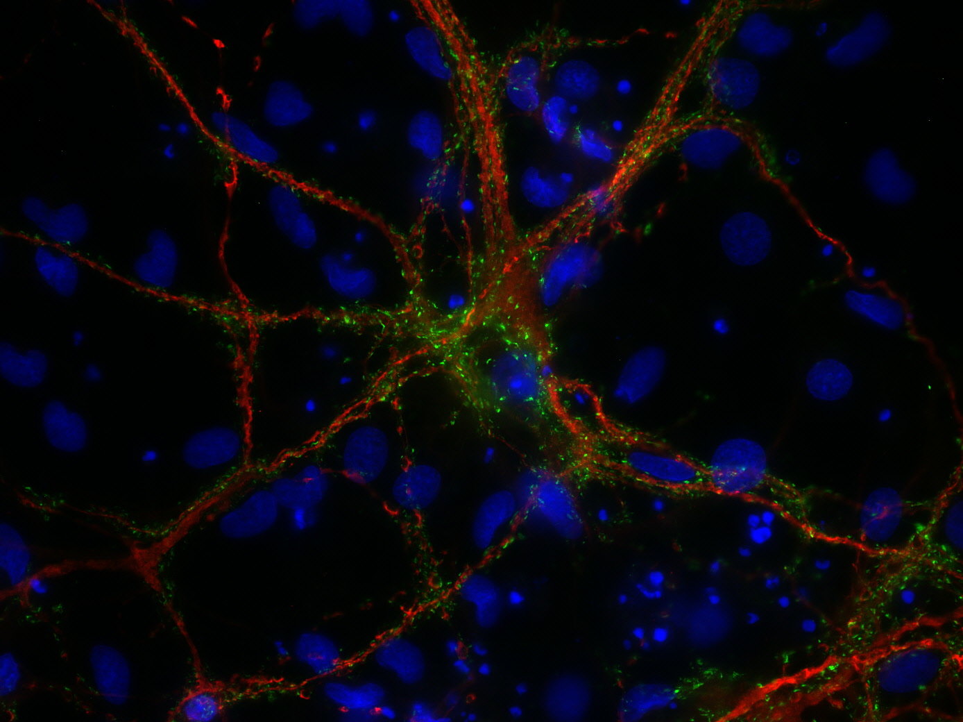
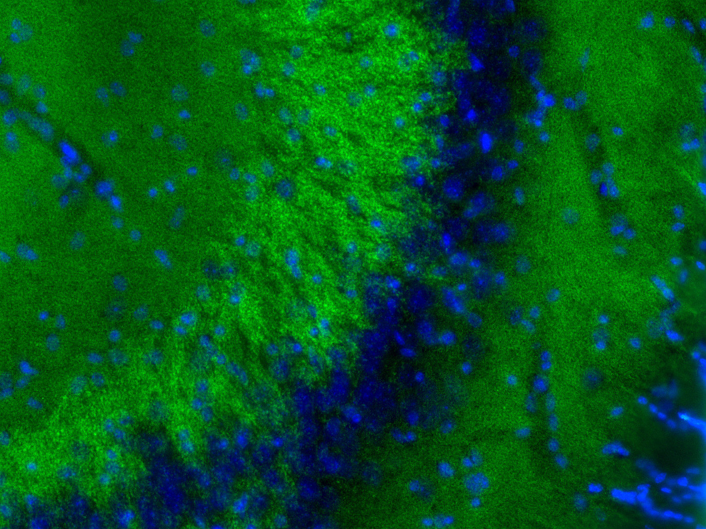
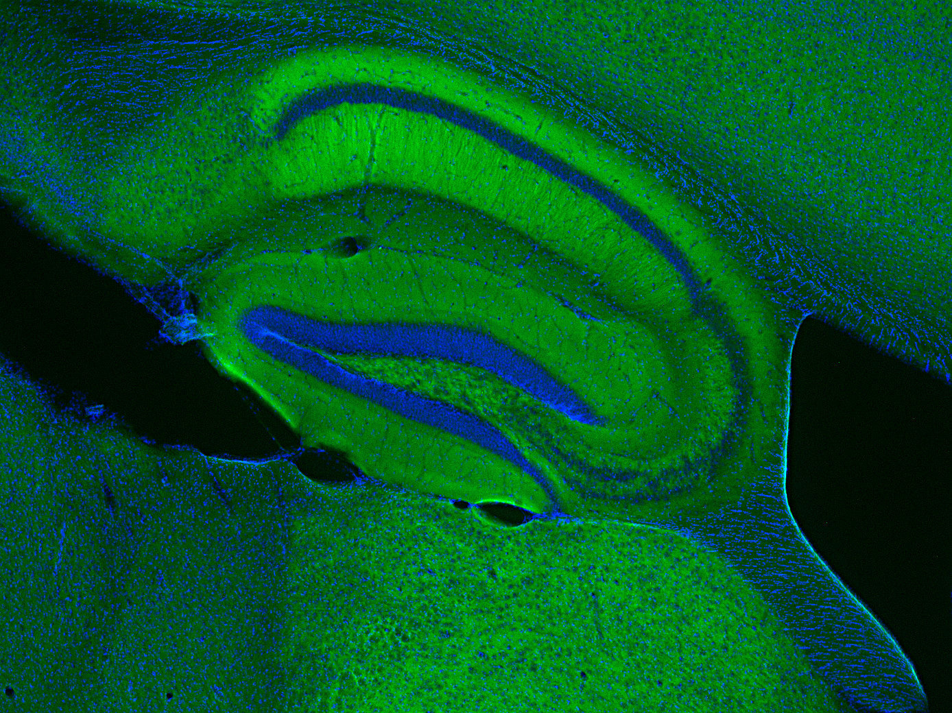
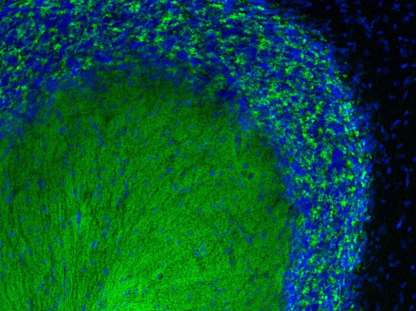
| Immunogen | Recombinant protein corresponding to AA 58 to 515 from rat VGLUT1 (UniProt Id: Q62634) |
| Reactivity |
Reacts with: rat (Q62634), mouse (Q3TXX4). Other species not tested yet. |
|
|
The vesicular glutamate transporter 1 VGLUT 1, also referred to as BNPI and SLC17A7, was originally identified as a brain specific phosphate transporter. Like the related VGLUT 2, VGLUT 1 is both necessary and sufficient for uptake and storage of glutamate and thus comprises the sole determinant for a glutamatergic phenotype. Both VGLUTs are different from the plasma membrane transporters in that they are driven by a proton electrochemical gradient across the vesicle membrane.
VGLUT 1 and VGLUT 2 show complementary expression patterns. Together, they are currently the best markers for glutamatergic nerve terminals and glutamatergic synapses.
Unlabeled variants and several modifications of sdAbs like biotin, fluorophore or DBCO conjugation are available.
In FluoTag®-X2 two fluorophore molecules are site-specifically coupled to each FluoTag molecule. Therefore, the reagent simultaneously targets two fluorophores to the protein of interest, which ensures up to two-fold („2X“)-brighter signals. Owing to the small size of the FluoTags, the distance between the target epitope and each fluorophore is ~ 3 nm.
In comparison to detection systems using conventional antibodies, FluoTag-X can thus improve the localization accuracy by 10-15 nm. Both features - superior brightness and precise fluorophore placement - render the FluoTag-X products excellent tools for all microscopy techniques.
Certificates
ISO 9001 2015 Quality Management System and Green Lab Platinum certification level for sustaining laboratory processes.


Newsletter
Sign up for our newsletter and get the latest updates and news.