Cat. No.: 398 117
Amount: 200 µl
Price:
$420.00


|

|
|
| Cat. No. 398 117 |
200 µl purified IgG, lyophilized. Albumin and azide were added for stabilization. For reconstitution add 200 µl H2O. Then aliquot and store at -20°C to -80°C until use. Antibodies should be stored at +4°C when still lyophilized. Do not freeze! |
| Applications | |
| Clone | 311H2 |
| Subtype | IgG2b (λ light chain) |
| Immunogen | Synthetic peptide corresponding to residues surrounding AA 1240 of mouse Ki67 (UniProt Id: E9PVX6) |
| Reactivity |
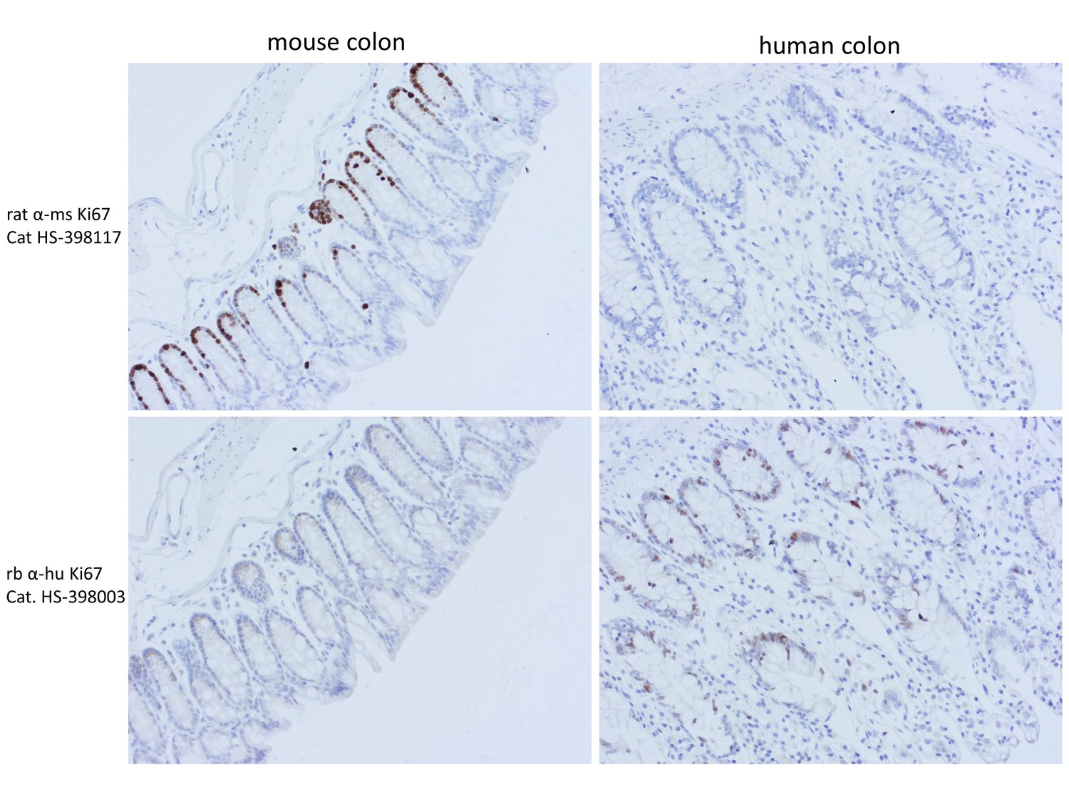
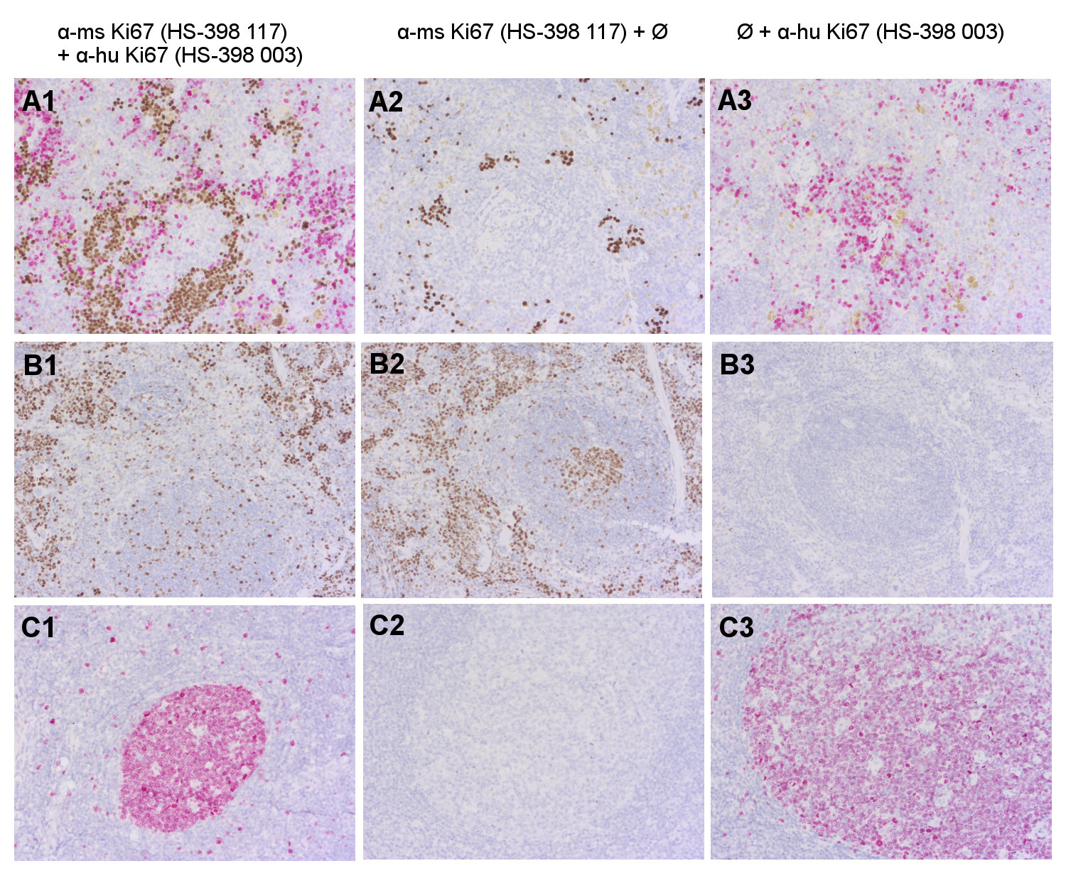
Reacts with: mouse (E9PVX6). No signal: human (P46013), rat (D4A0Y6), chicken, zebrafish. Other species not tested yet. |
| Remarks |
IHC: Antigen retrieval with citrate buffer pH 6 is required. |
| Data sheet | Data Sheet 398_117 |
|
|
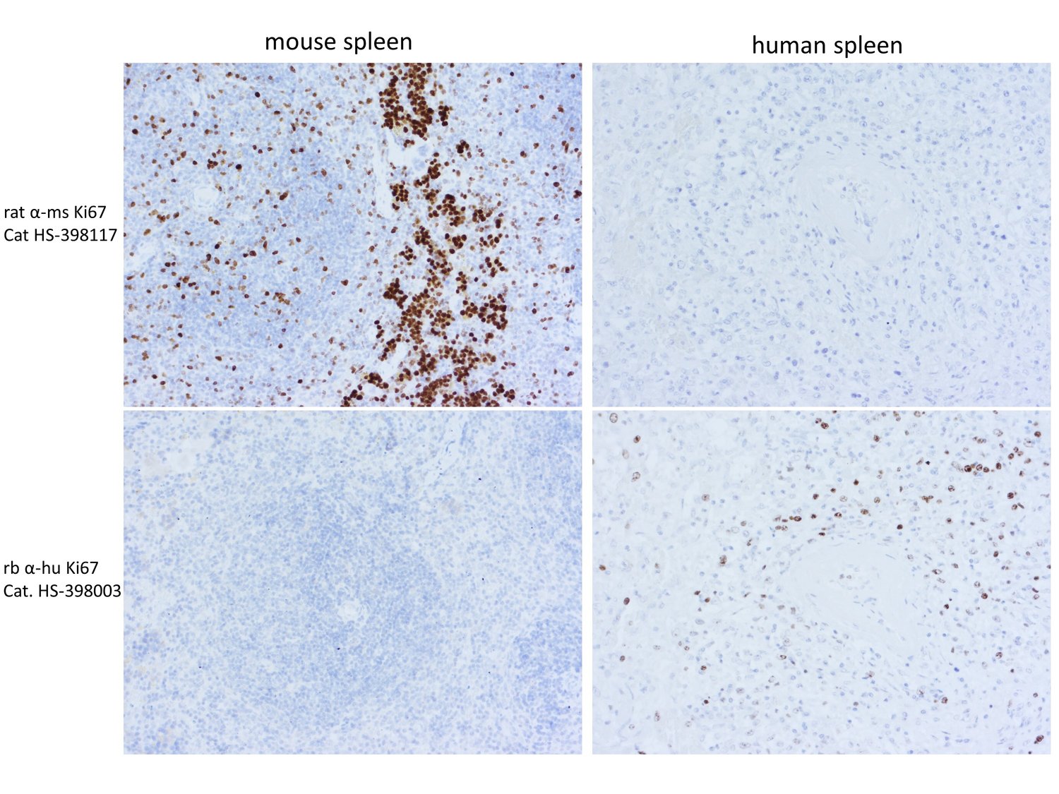
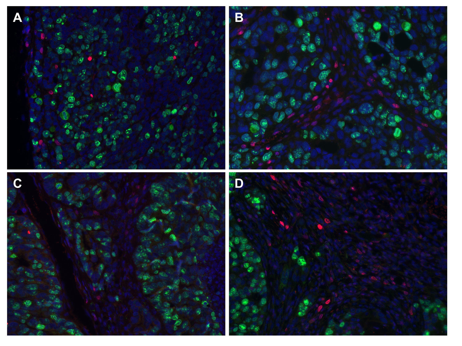
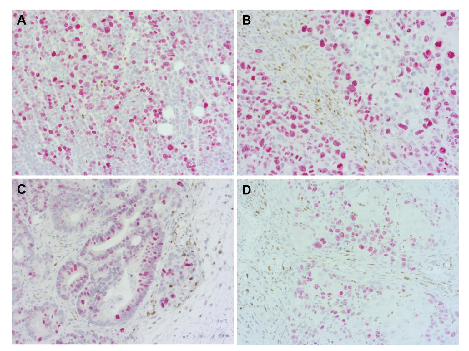
Species-specific detection of proliferation antigen-Ki67 in murine but not human FFPE spleen
Ki-67 is a nuclear protein which is widely used as a marker of cell proliferation. Its expression is closely associated with the cell cycle: Ki-67 is present in actively cycling cells during the G1, S, G2, and M phases but is generally absent in quiescent cells in the G0 phase. At the cellular level, Ki-67 is predominantly localized in the nucleus during interphase and redistributes to the periphery of condensed chromosomes during mitosis (1,2).
Immunohistochemical staining for Ki-67 is a simple and reproducible method to determine the tumor proliferative index and is a predictive and prognostic biomarker in certain types of human cancer, such as breast cancer, gastric cancer or prostate cancer. In breast cancer higher Ki-67 levels have been associated with an increased risk of disease recurrence (1,3). However, Ki-67 expression is not specific to malignant cells, as it is also present in normally proliferating tissues and activated lymphocytes.
Certificates
ISO 9001 2015 Quality Management System and Green Lab Platinum certification level for sustaining laboratory processes.


Newsletter
Sign up for our newsletter and get the latest updates and news.