Cat. No.: 538 017
Amount: 100 µg
Price:
$420.00
|
|

|
|
| Cat. No. 538 017 |
100 µg purified IgG, lyophilized. Albumin and azide were added for stabilization. For reconstitution add 100 µl H2O to get a 1mg/ml solution in PBS. Then aliquot and store at -20°C to -80°C until use. Antibodies should be stored at +4°C when still lyophilized. Do not freeze! |
| Applications | |
| Clone | SY-244C1 |
| Subtype | IgG2b (κ light chain) |
| Immunogen | Full-length recombinant mouse Lymphocyte antigen 6C1 protein (UniProt Id: P0CW02) |
| Specificity | Specific for mouse Ly6C; no cross-reactivity with mouse Ly6G |
| Remarks |
ICC: 4% formaldehyde/PFA fixation is recommended. |
| Data sheet | Datasheet 538_017 |
|
|
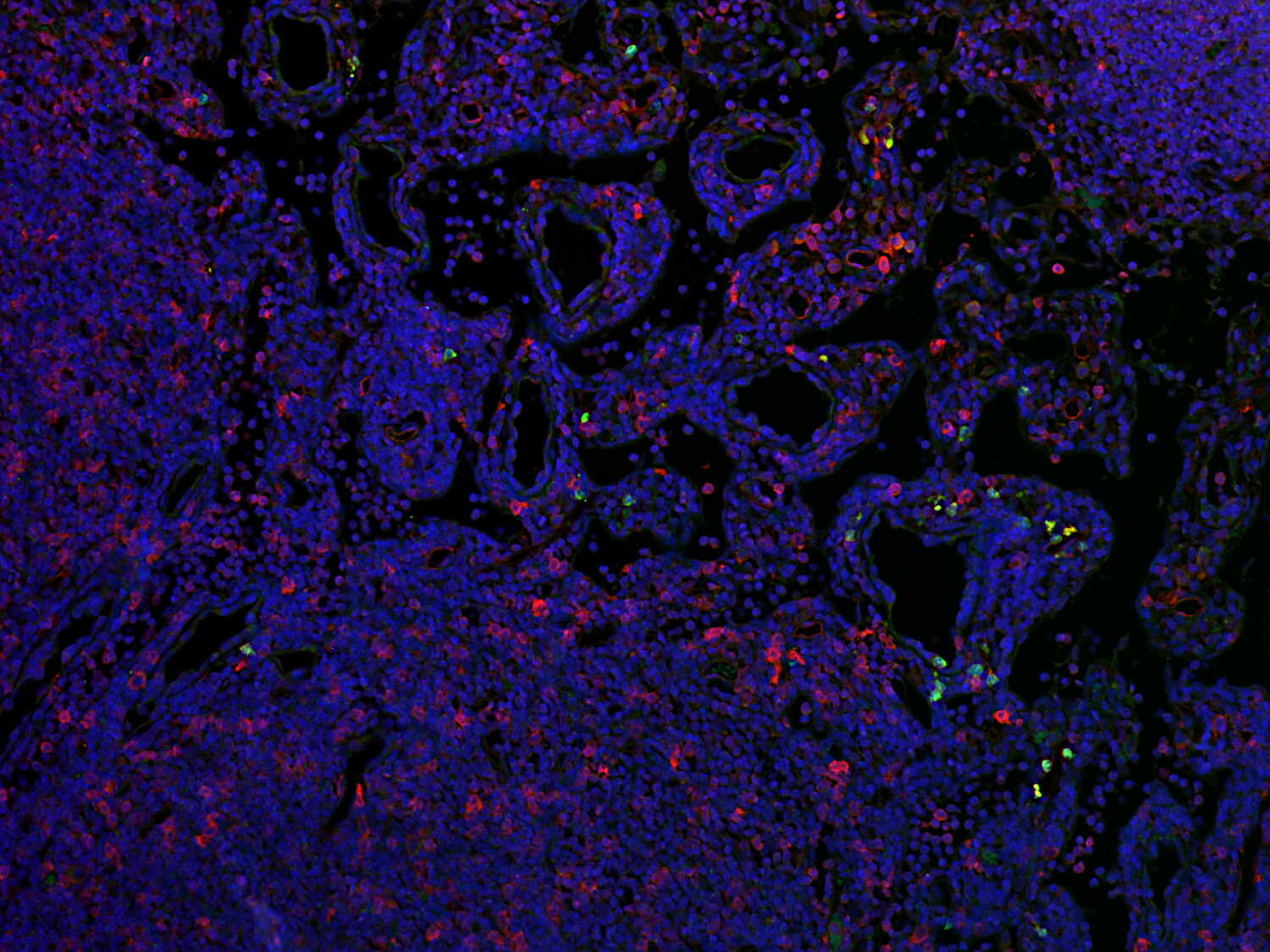
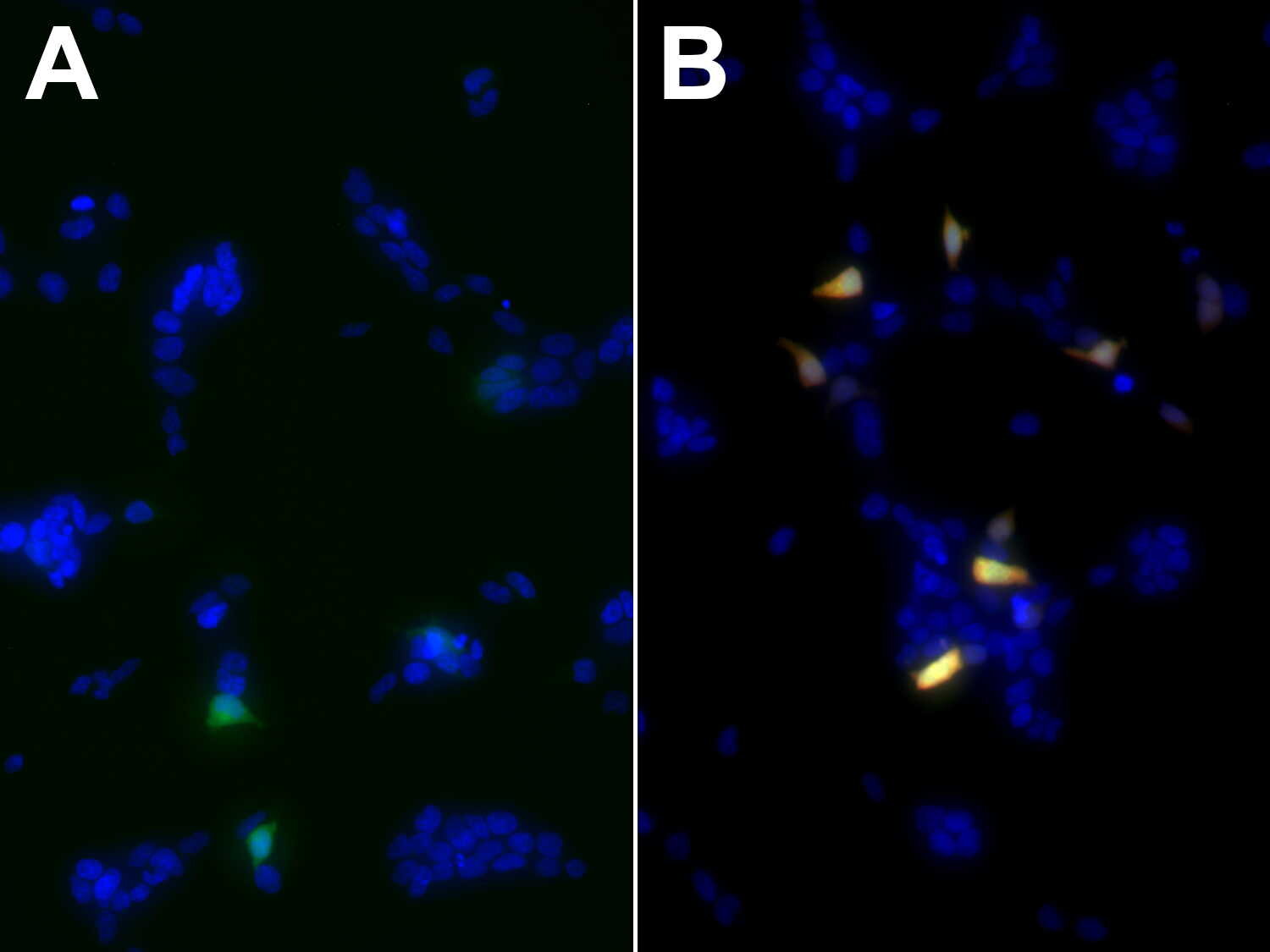
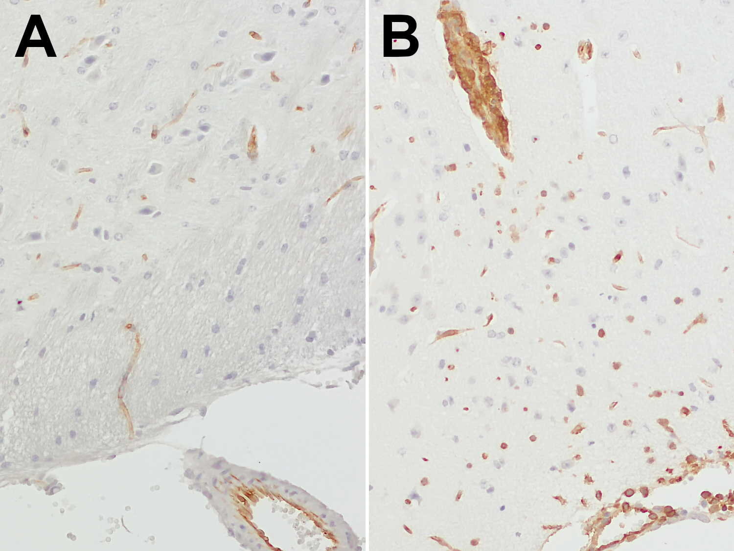
Ly6C (red) and Ly6G (green) distinguish different myeloid populations in mouse lymph node
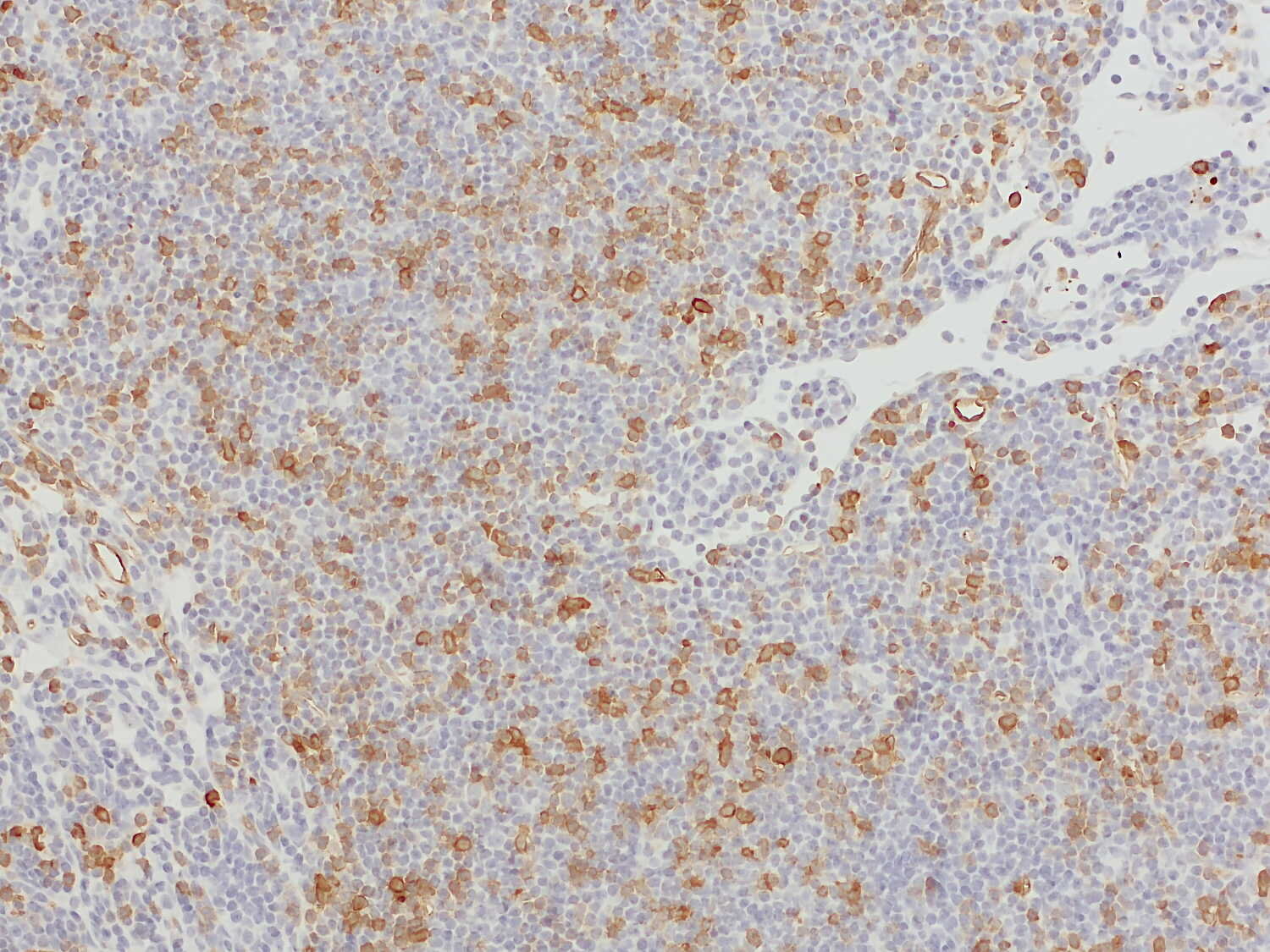
Ly6C1 and Ly6C2 (Lymphocyte antigen 6 complex, locus C 1 and 2) are two highly homologous proteins, that are collectively known as Ly6C. Ly6C is a 14-kD glycosylphosphatidylinositol (GPI)-anchored protein that plays a crucial role in immune regulation and cell adhesion. It contains a single Cys-rich motif that generates the LU domain structure characteristic of the Ly6 family (1). Ly6C is commonly used as a monocyte/macrophage cell differentiation antigen. Ly6C high pro-inflammatory macrophages develop from recruited classical Ly6C high monocytes during inflammation and are then converted into Ly6C low macrophages, that are more involved in tissue repair and resolution of inflammation (2).
In the steady state, tissue-resident macrophages, such as microglia, Langerhans cells, and Kupffer cells, exhibit a F4/80highLy6Clow phenotype (2). Ly6C is primarily observed on monocytes, dendritic cells, and a subset of T cells (3,4). Ly6C is also expressed on brain vessel endothelial cells (5) and in the retinal vascular plexuses (6).
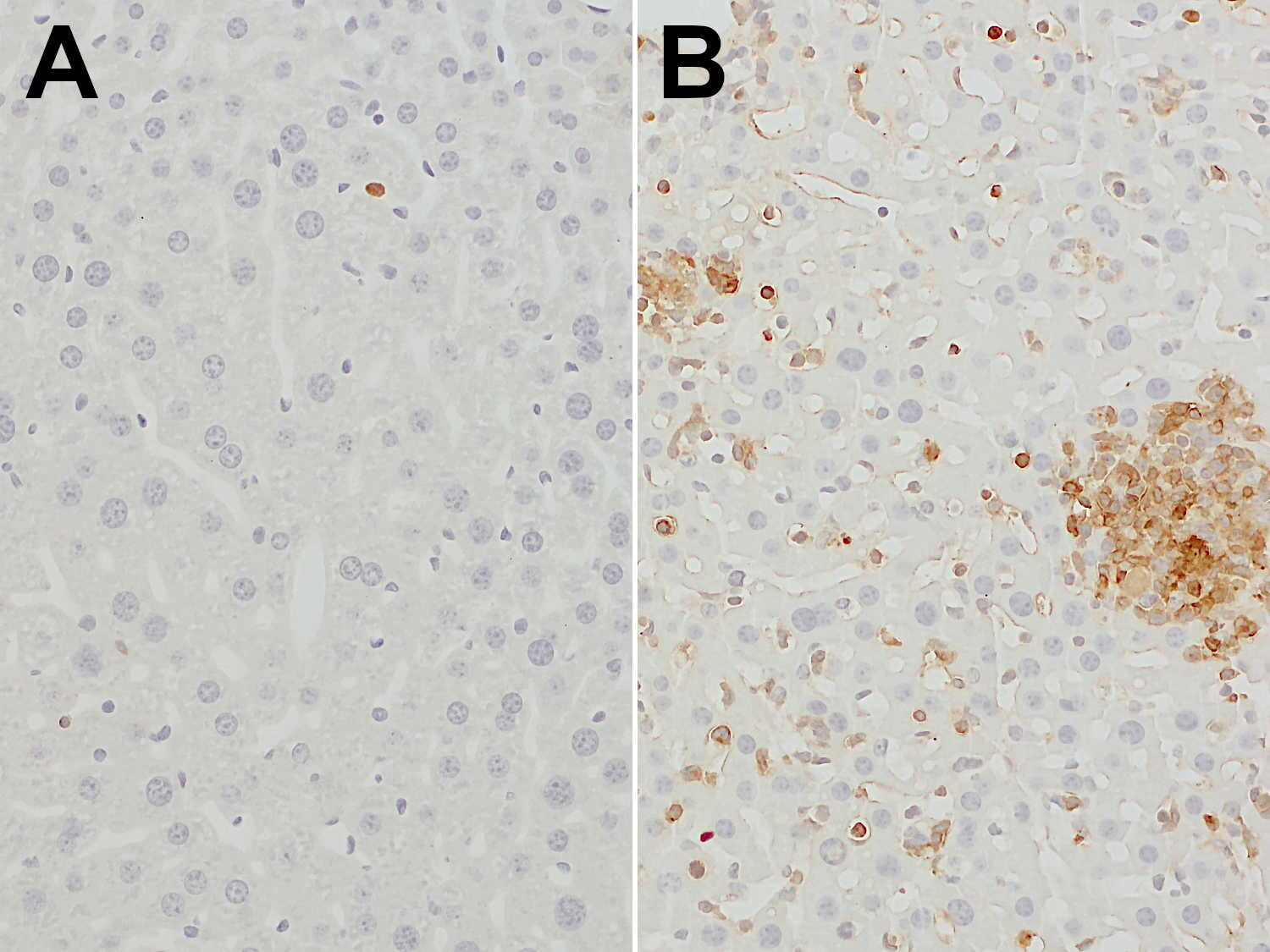
In disease contexts, particularly in chronic inflammatory conditions, cancer, and autoimmune disorders, increased levels of Ly6C high monocytes have been linked to exacerbated inflammation and tissue damage (7).
Certificates
ISO 9001 2015 Quality Management System and Green Lab Platinum certification level for sustaining laboratory processes.


Newsletter
Sign up for our newsletter and get the latest updates and news.