Cat. No.: 397 017
Amount: 200 µl
Price:
$420.00


|

|
|
| Cat. No. 397 017 |
200 µl purified IgG, lyophilized. Albumin and azide were added for stabilization. For reconstitution add 200 µl H2O. Then aliquot and store at -20°C to -80°C until use. Antibodies should be stored at +4°C when still lyophilized. Do not freeze! |
| Applications | |
| Clone | 167B3 |
| Subtype | IgG2a (κ light chain) |
| Immunogen | Synthetic peptide corresponding to residues surrounding AA36 of mouse F4/80 (UniProt Id: Q61549) |
| Reactivity |
Reacts with: mouse (Q61549). No signal: rat. Other species not tested yet. |
| Remarks |
IHC-Fr: The following fixatives are possible: methanol, 4% formaldehyde/PFA. |
| Data sheet | Data Sheet 397_017 |
|
|
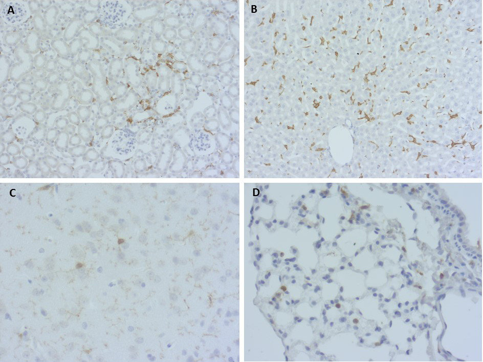
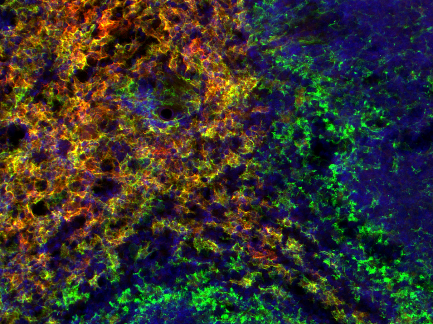
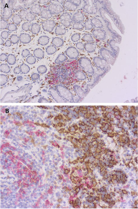
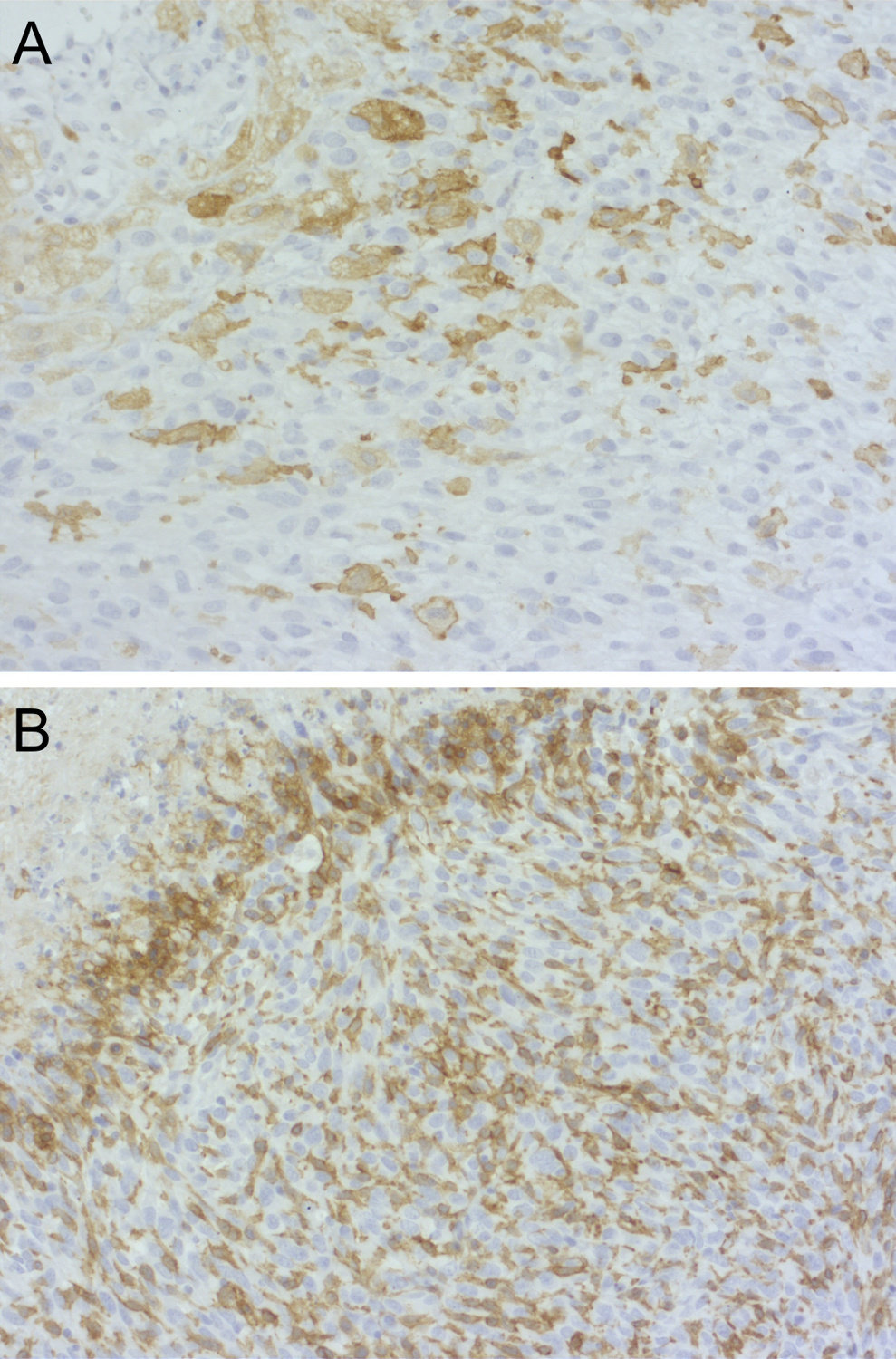
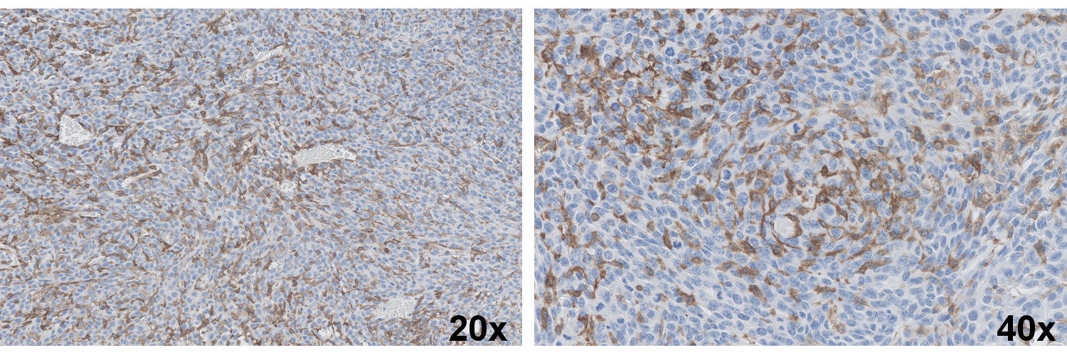
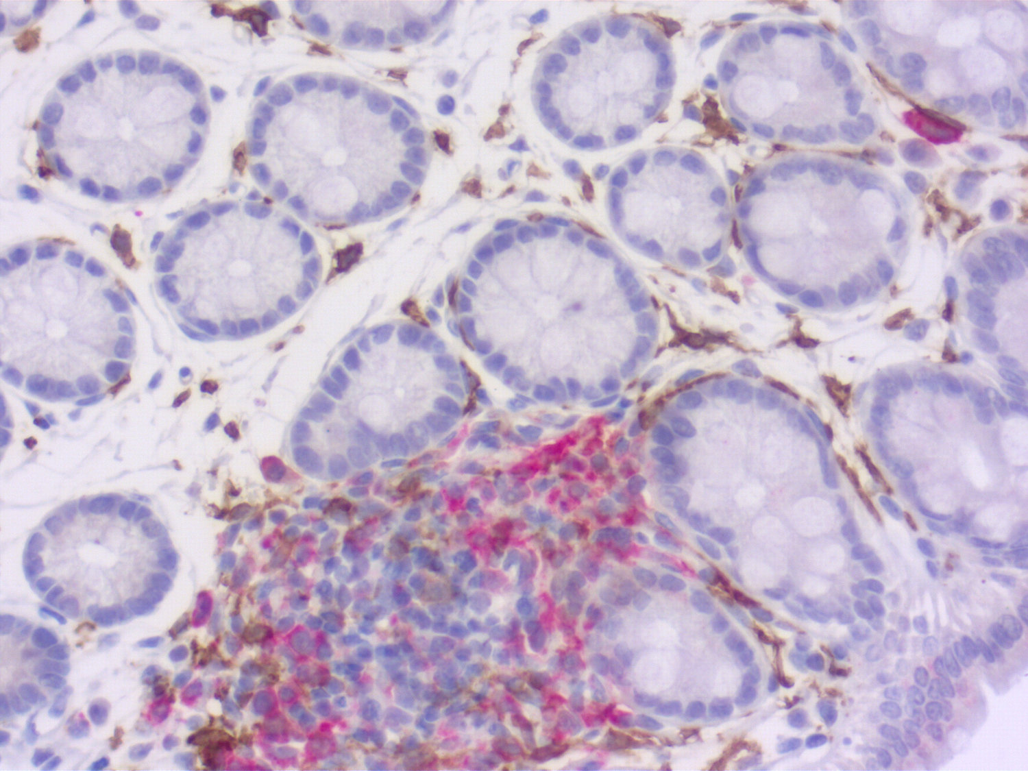
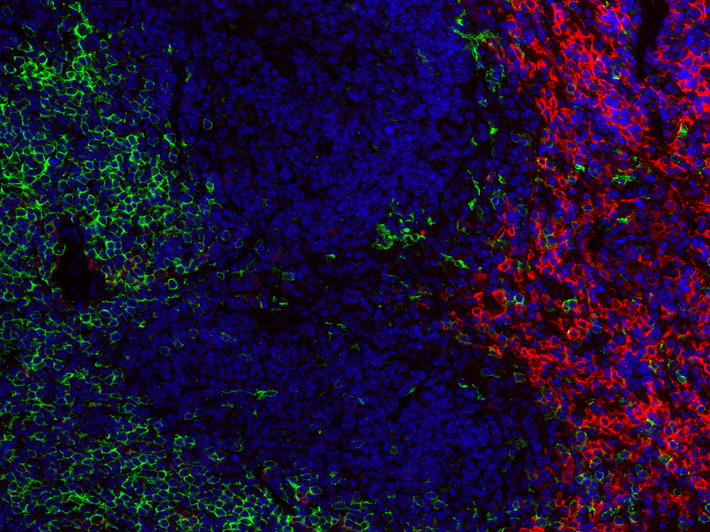
F4/80 positive cells in murine FFPE tissues
F4/80, also known as EMR1 or ADGRE1, is a seven-transmembrane adhesion G protein-coupled receptor that is widely regarded as a canonical surface marker of tissue-resident macrophages in the mouse. It is a member of the ADGRE (EGF-TM7) family and is highly expressed by splenic red pulp macrophages, hepatic Kupffer cells, epidermal Langerhans cells, microglia, and intestinal macrophages, whereas its expression is largely absent from circulating monocytes and most dendritic cell subsets (1,2).
Within the spleen, F4/80⁺ red pulp macrophages are critically involved in the clearance of senescent erythrocytes and the recycling of iron through heme oxygenase-1, thereby contributing to the maintenance of iron homeostasis during blood-borne infections such as malaria (1). Beyond its value as a phenotypic marker, F4/80 has also been implicated in immunoregulatory processes. Notably, F4/80-deficient mice fail to generate the antigen-specific CD8⁺ regulatory T cells required for peripheral tolerance, and administration of anti-F4/80 antibodies inhibits the induction of anterior chamber-associated immune deviation (3).
In inflammatory bowel disease models, an F4/80⁺Ly6C^high macrophage subset expands during dextran sulfate sodium (DSS)-induced colitis and promotes tumor initiation through the secretion of pro-inflammatory cytokines, thereby linking this population to the pathogenesis of colitis-associated cancer (4). Across mammalian species, expression of the F4/80 orthologue ADGRE1 is conserved in monocytes and macrophages; however, the extracellular EGF-like domains are highly variable between species, suggesting a potential role in pathogen recognition (5).
For more information on protein expression pattern, please refer to the overview image in our SYSY Antibodies ATLAS.
Certificates
ISO 9001 2015 Quality Management System and Green Lab Platinum certification level for sustaining laboratory processes.


Newsletter
Sign up for our newsletter and get the latest updates and news.