Ca2+ channel N-type alpha-1B antibody - 152 313 K.D.

Rabbit polyclonal purified antibody
Cat. No.: 152 313
Amount: 50 µg
Price: $380.00

Cat. No. 152 313 50 µg specific antibody, lyophilized. Affinity purified with the immunogen. Albumin and azide were added for stabilization. For reconstitution add 50 µl H2O to get a 1mg/ml solution in PBS. Then aliquot and store at -20°C to -80°C until use.
Antibodies should be stored at +4°C when still lyophilized. Do not freeze!
Applications
 
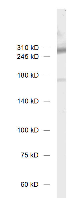
WB: 1 : 1000 (AP staining) gallery
IP: not tested yet
ICC: external data (see remarks)
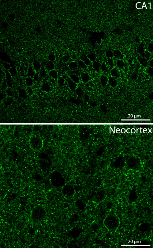
IHC: external data (see remarks) gallery
IHC-P (FFPE): not tested yet
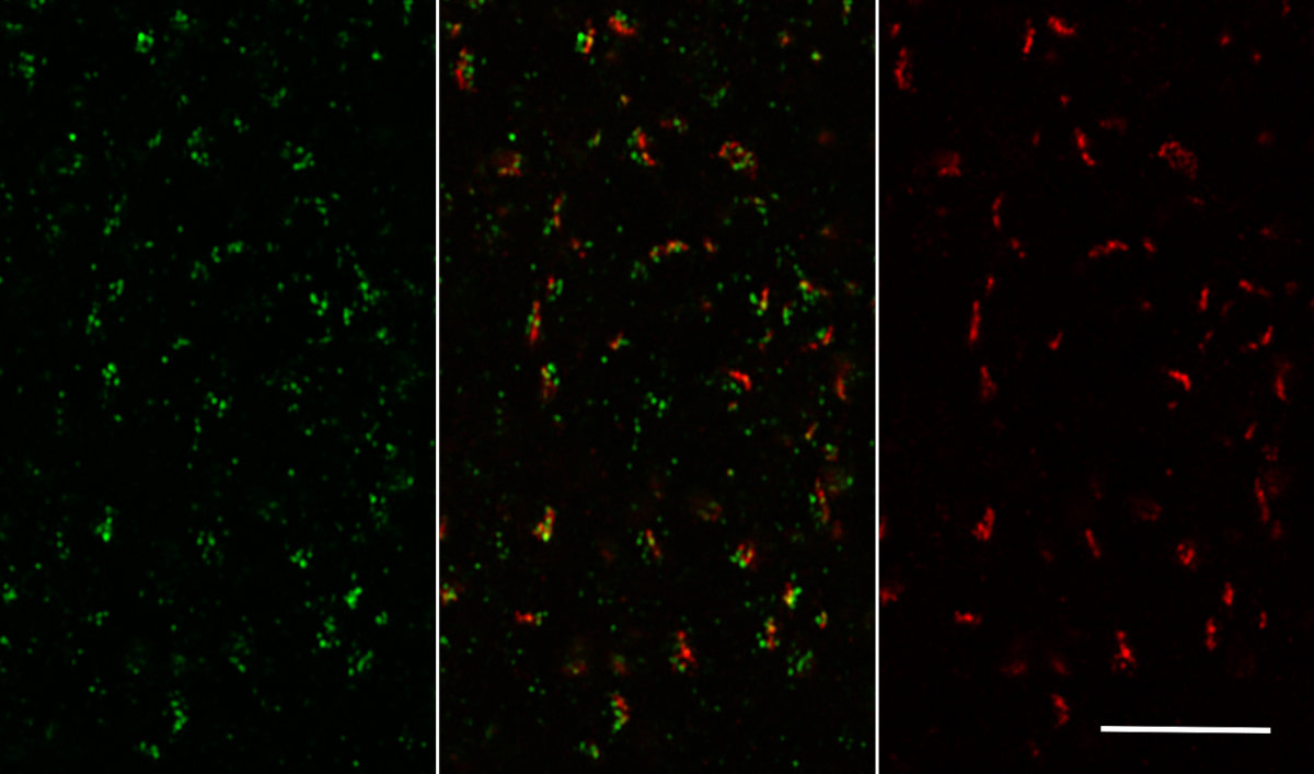
ExM: external data (see remarks) gallery
EM: not tested yet

Western blot (WB); separation of proteins by PAGE and subsequent transfer to a membrane. Detection of target molecules is carried out with antibodies. Some antibodies require special sample preparation steps. For details, please refer to the “Remarks” section.

Immunoprecipitation (IP); Immunoisolation or pulldown of a target molecule using an antibody. For details and product specific hints, please refer to the ”Remarks” section.

Immunocytochemistry (ICC) on 4% PFA fixed cells. Immunoreactivity is usually revealed by fluorescence. Some antibodies require special fixation methods. For details, please refer to the “Remarks” section.

Immunohistochemistry (IHC) on 4% PFA perfusion fixed tissue with 24h PFA post fixation. Immunoreactivity is usually revealed by fluorescence or a chromogenic substrate. Some antibodies require special fixation methods or antigen retrieval steps. For details, please refer to the ”Remarks” section.

Immunohistochemistry (IHC-P) of formalin fixed, paraffin embedded (FFPE) tissue (some antibodies require special antigen retrieval steps, please refer to the ”Remarks” section). Immunoreactivity is usually revealed by fluorescence or a chromogenic substrate.

Expansion Microscopy (ExM) is a sample preparation and imaging method which employs a dense interconnected web of swellable polymer within a biological specimen. A significantly higher effective resolution can be achieved with a standard microscope setup. For additional experimental details, please refer to the Remarks section.

Electron microscopy (EM) is a microscopy technique that detects the scatter of electrons through thin tissue sections. In Immuno-EM the antigen is usually revealed by colloidal gold conjugated secondary antibodies linking electron dense structures to antigen-bound primary antibodies.

Immunogen Recombinant protein corresponding to AA 2056 to 2336 from rat Ca2+ channel N-type α-1B (Cav2.2) (UniProt Id: Q02294)
Reactivity Reacts with: rat (Q02294), mouse (O55017).
Other species not tested yet.
Specificity K.D. validated PubMed: 29198756
Matching control protein/peptide 152-3P
Remarks

ICC: This antibody has been successfully used and published for this application by customers (see application-specific references). It is not compatible with our standard protocols.
IHC: This antibody has been successfully applied for this method by our customers using mild fixation (1% PFA at pH 6) according to Lorincz and Nusser 2010 (see gallery). It has not been validated using our standard protocol.
ExM: This antibody has been successfully applied and published for this method by customers (see application-specific references).

Data sheet Datasheet 152_313

References for Ca2+ channel N-type alpha-1B - 152 313

Physiological super-resolution in situ recording reveals kinetic changes of sympathetic norepinephrine release in heart failure.
Liu B, Jiang X, Guo S, Kang X, Lü J, Wang S, Yao W, Ou X, Yan J, Li T, Gu J, et al.
Proceedings of the National Academy of Sciences of the United States of America (2026) 12318: e2519828123. 152 313 WB; tested species: mouse
CaV2.2 Gates Calcium-Independent but Voltage-Dependent Secretion in Mammalian Sensory Neurons.
Chai Z, Wang C, Huang R, Wang Y, Zhang X, Wu Q, Wang Y, Wu X, Zheng L, Zhang C, Guo W, et al.
Neuron (2017) 966: 1317-1326.e4. 152 313 WB, ICC; KD verified; tested species: rat
Selected Ionotropic Receptors and Voltage-Gated Ion Channels: More Functional Competence for Human Induced Pluripotent Stem Cell (iPSC)-Derived Nociceptors.
Schoepf CL, Zeidler M, Spiecker L, Kern G, Lechner J, Kummer KK, Kress M
Brain sciences (2020) 106: . 152 313 ICC; tested species: human,mouse
CaV2.2 Gates Calcium-Independent but Voltage-Dependent Secretion in Mammalian Sensory Neurons.
Chai Z, Wang C, Huang R, Wang Y, Zhang X, Wu Q, Wang Y, Wu X, Zheng L, Zhang C, Guo W, et al.
Neuron (2017) 966: 1317-1326.e4. 152 313 WB, ICC; KD verified; tested species: rat
Cat. No.: 152 313
Amount: 50 µg
Price: $380.00
Physiological super-resolution in situ recording reveals kinetic changes of sympathetic norepinephrine release in heart failure.
Liu B, Jiang X, Guo S, Kang X, Lü J, Wang S, Yao W, Ou X, Yan J, Li T, Gu J, et al.
Proceedings of the National Academy of Sciences of the United States of America (2026) 12318: e2519828123. 152 313 WB; tested species: mouse
CaV2.2 Gates Calcium-Independent but Voltage-Dependent Secretion in Mammalian Sensory Neurons.
Chai Z, Wang C, Huang R, Wang Y, Zhang X, Wu Q, Wang Y, Wu X, Zheng L, Zhang C, Guo W, et al.
Neuron (2017) 966: 1317-1326.e4. 152 313 WB, ICC; KD verified; tested species: rat
Selected Ionotropic Receptors and Voltage-Gated Ion Channels: More Functional Competence for Human Induced Pluripotent Stem Cell (iPSC)-Derived Nociceptors.
Schoepf CL, Zeidler M, Spiecker L, Kern G, Lechner J, Kummer KK, Kress M
Brain sciences (2020) 106: . 152 313 ICC; tested species: human,mouse
CaV2.2 Gates Calcium-Independent but Voltage-Dependent Secretion in Mammalian Sensory Neurons.
Chai Z, Wang C, Huang R, Wang Y, Zhang X, Wu Q, Wang Y, Wu X, Zheng L, Zhang C, Guo W, et al.
Neuron (2017) 966: 1317-1326.e4. 152 313 WB, ICC; KD verified; tested species: rat
Background

Voltage gated calcium channels (VGCCs), also referred to as voltage sensitive calcium channels (VSCCs), are present in most excitable cells. They mediate the influx of Ca2+ ions into the cell and trigger the release of neurotransmitters or hormons but are also involved in other calcium dependent processes like metabolism, cell proliferation and cell death.
VGCCs are composed of four subunits (α-1, α-2, β and δ) in a 1:1:1:1 ratio. The α-1A isoform occurs in VGCCs of the P/Q-type while isoform α-1B is found in the N-type. Both belong to the high voltage activated group (hva).

Protocols
WB
ICC
  • see remarks
IHC
  • see remarks

Certificates

ISO 9001 2015 Quality Management System and Green Lab Platinum certification level for sustaining laboratory processes.

Newsletter

Sign up for our newsletter and get the latest updates and news.
 

Please provide a valid E-Mail-Address
Please provide a valid First name
Please provide a valid Last name