Cat. No.: 119 008
Amount: 50 µg
Price:
$420.00
|
|

|
|
| Cat. No. 119 008 |
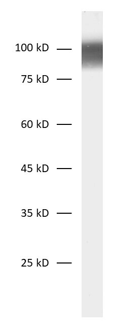
50 µg purified recombinant IgG, lyophilized. Albumin and azide were added for stabilization. For reconstitution add 50 µl H2O to get a 1mg/ml solution in PBS. Then aliquot and store at -20°C to -80°C until use. Antibodies should be stored at +4°C when still lyophilized. Do not freeze! |
| Applications | |
| Clone | Rb188D1 |
| Subtype | IgG1 (κ light chain) |
| Immunogen | Synthetic peptide corresponding to residues near the amino terminus of human SV2 A (UniProt Id: Q7L0J3) |
| Reactivity |
Reacts with: mouse (Q9JIS5), rat (Q02563). Other species not tested yet. Predicted to cross-react with human (Q7L0J3) due to high sequence homology. |
| Remarks |
This antibody is a chimeric antibody based on the monoclonal mouse antibody SY-188D1. The constant regions of the heavy and light chains have been replaced with rabbit specific sequences. The antibody can therefore be used with standard anti-rabbit secondary reagents. The antibody has been expressed in mammalian cells. |
| Data sheet | Datasheet 119_008 |
|
|
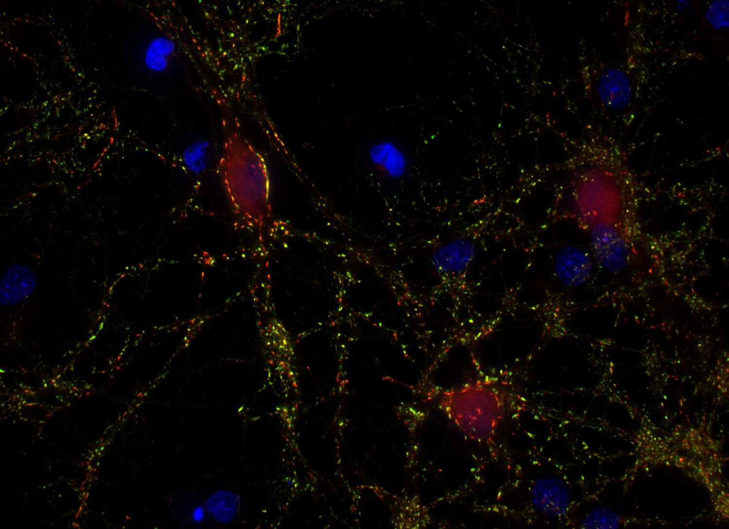
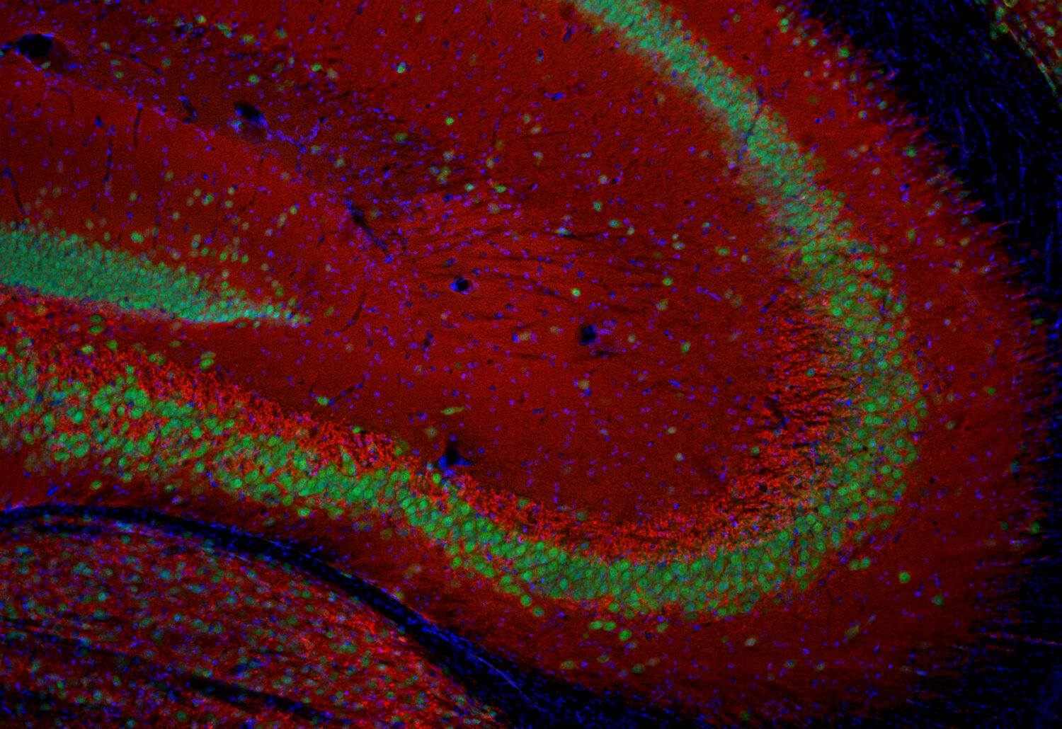
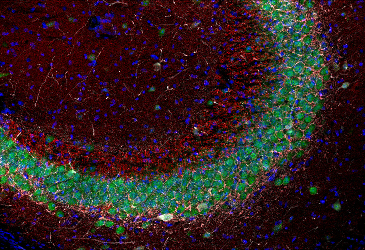
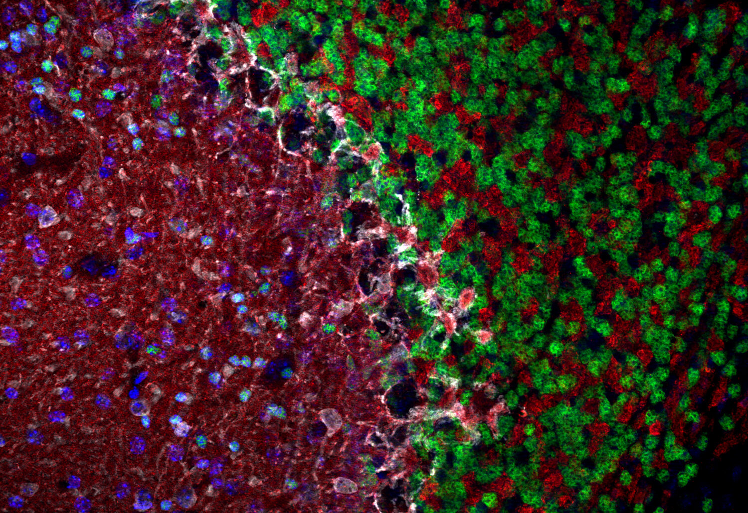
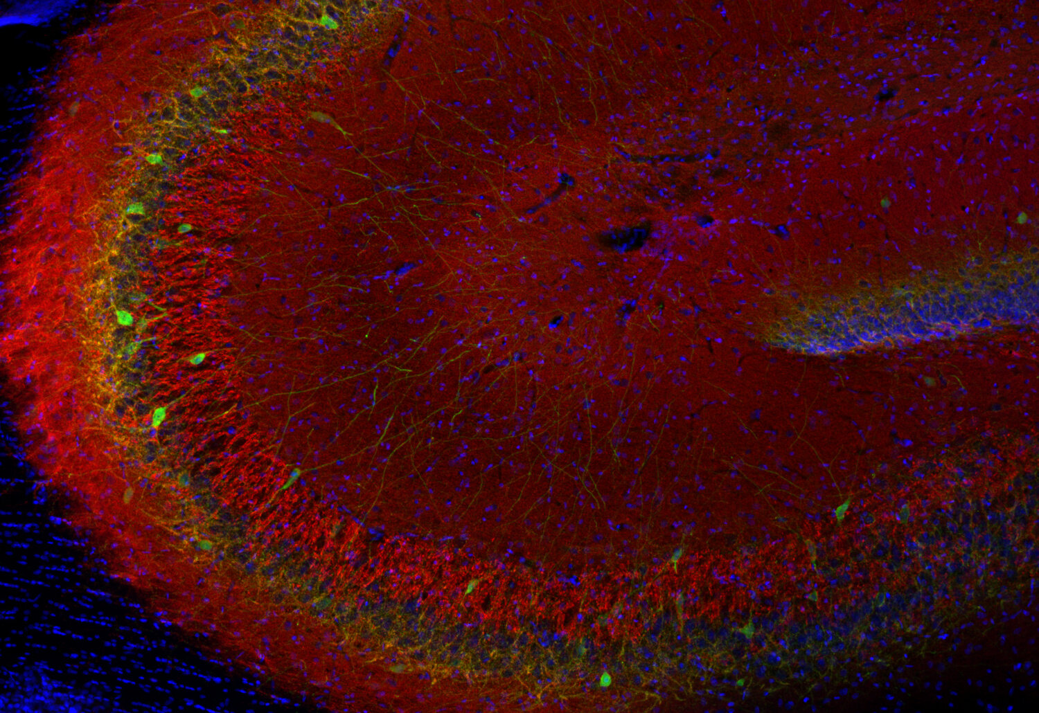
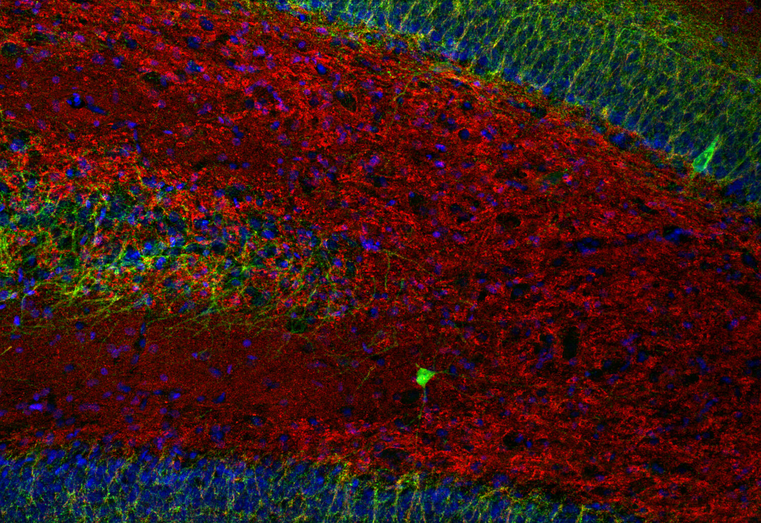
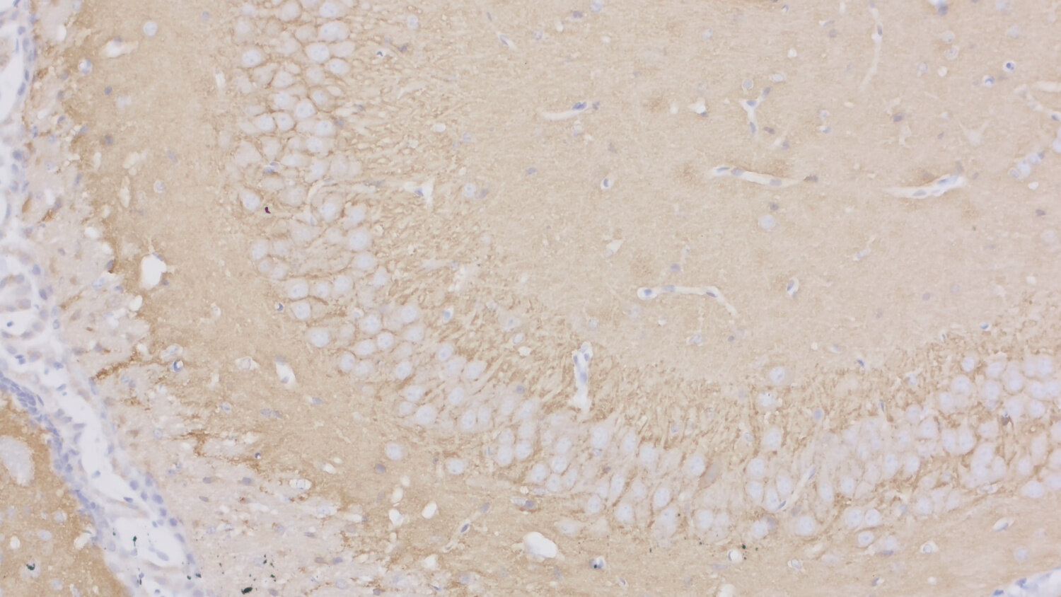
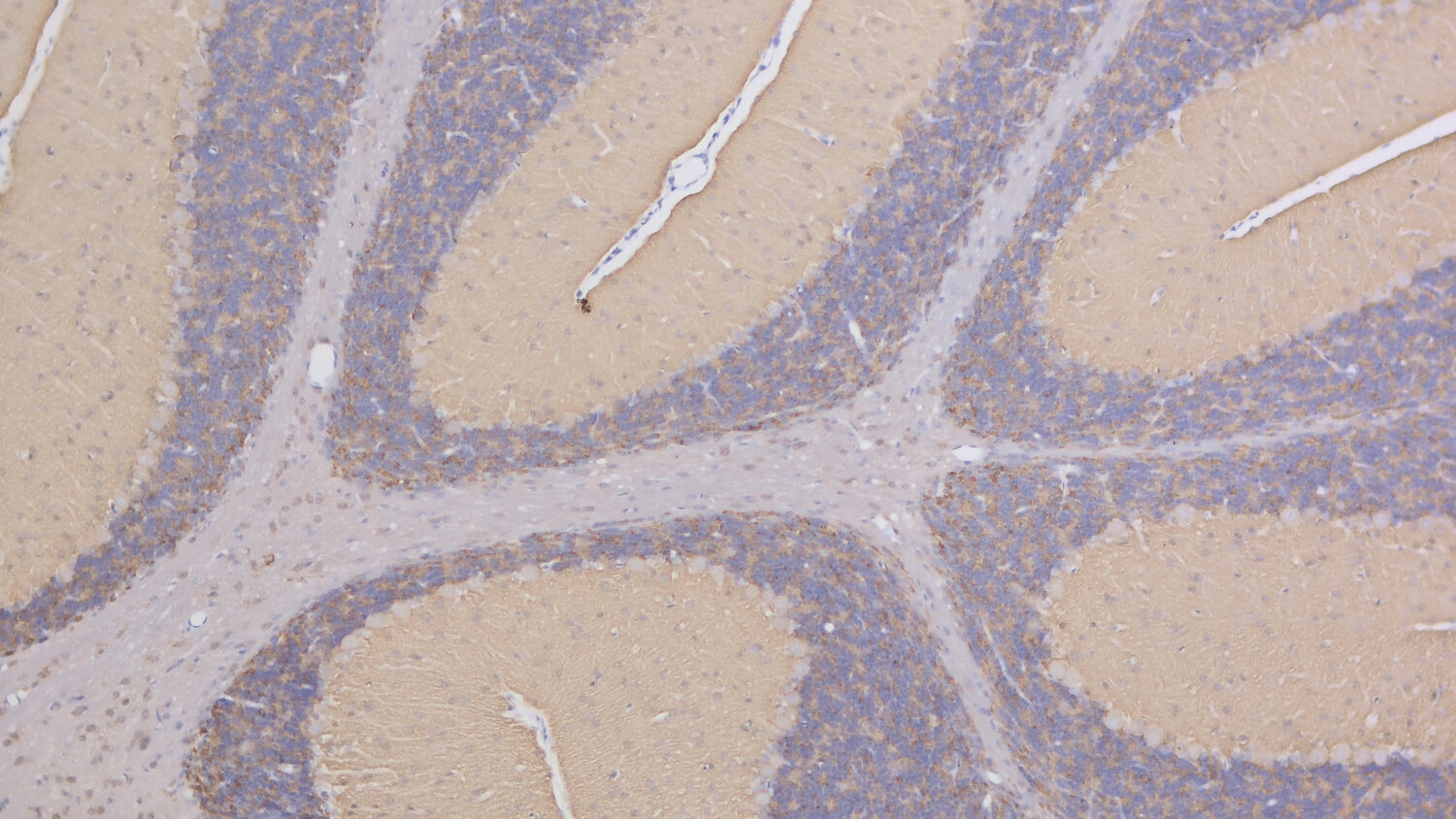
SV2s (Synaptic Vesicle Protein 2) are integral membrane glycoproteins present in synaptic vesicles. They have 12 transmembrane domains predicted by sequence analysis (1). There are three characterized isoforms, SV2 A, SV2 B and SV2 C that are similar in structure but show different expression patterns. SV2 A is expressed ubiquitously throughout the brain and plays a crucial role in modulating synaptic transmission by regulating the expression and trafficking of synaptotagmin, a key calcium sensor in neurotransmitter release (1).
SV2 B has a more restricted distribution with varying degrees of coexpression with SV2 A and is predominantly found in the cortex and hippocampus (2). SV2 C is more closely related to SV2 A but shows a very restricted expression pattern. The highest expression levels were observed in phylogenetically old brain areas like pallidum, the midbrain and the olfactory bulb (3).
SV2 expression has also been observed in other non-neuronal organs. In kidney it localizes to podocytes and is essential for the integrity of the glomerular filtration barrier (4).
For more information on protein expression pattern, please refer to the overview image in our SYSY Antibodies ATLAS.
Certificates
ISO 9001 2015 Quality Management System and Green Lab Platinum certification level for sustaining laboratory processes.


Newsletter
Sign up for our newsletter and get the latest updates and news.