Cat. No.: 104 002
Amount: 200 µl
Price:
$360.00
|
|

|
|
| Cat. No. 104 002 |
200 µl antiserum, lyophilized. For reconstitution add 200 µl H2O, then aliquot and store at -20°C until use. Antibodies should be stored at +4°C when still lyophilized. Do not freeze! |
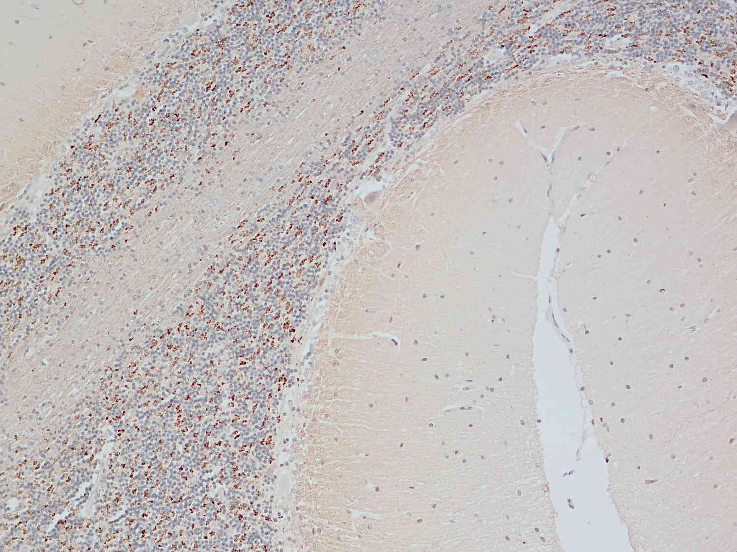
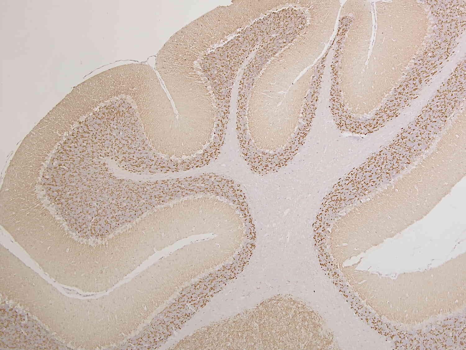
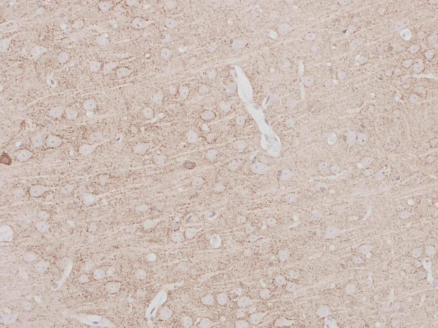
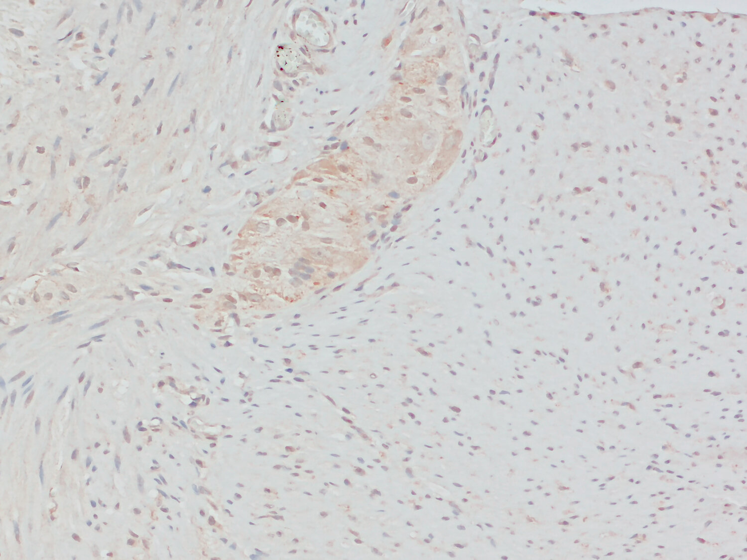
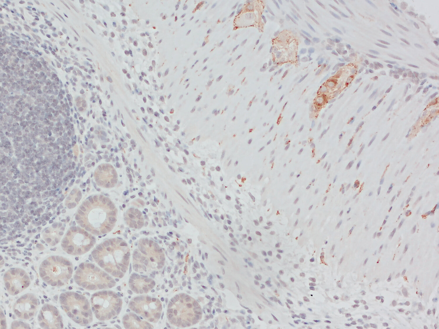
| Applications | |
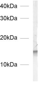
| Immunogen | Synthetic peptide corresponding to AA 2 to 14 from rat Synaptobrevin1 (UniProt Id: Q63666) |
| Reactivity |
Reacts with: human (P23763), rat (Q63666), mouse (Q62442), monkey, hamster, human (P23763). No signal: chicken, cat. Other species not tested yet. |
| Specificity | K.D. validated PubMed: 29621484 |
| Matching control protein/peptide | 104-0P |
| Remarks |
EM: This antibody has been successfully applied and published for this method by customers (see application-specific references). |
| Data sheet | Datasheet 104_002 |
|
|
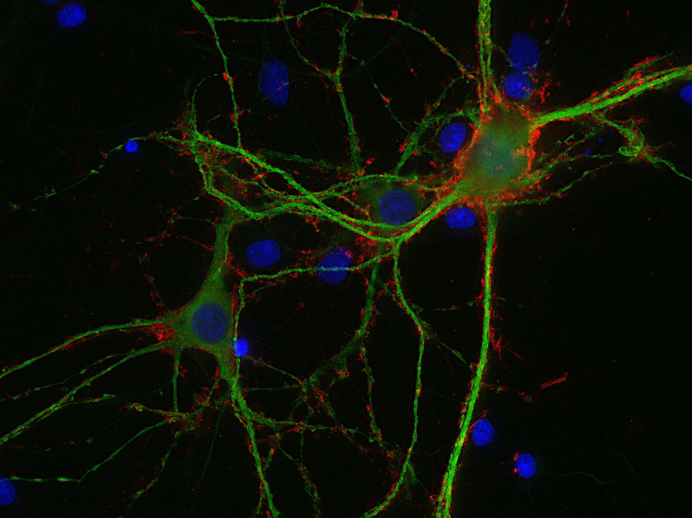
Synaptobrevins, also known as vesicle-associated membrane proteins (VAMPs), are predominantly expressed in the nervous system and are classified within the brevin subfamily of the SNARE (Soluble NSF Attachment Protein Receptor) protein superfamily. Brevins are small integral transmembrane proteins characterized by a central SNARE motif, an N-terminal cytoplasmic domain, and a C-terminal transmembrane domain. As crucial components of the SNARE machinery, these proteins play an essential role in vesicular transport and membrane fusion processes within cells (1, 2, 3).
In addition to synaptobrevins, the brevin family includes other tissue-specific members such as cellubrevin (VAMP3), myobrevin (VAMP5), and endobrevin (VAMP8), which are expressed in various non-neuronal tissues (4, 5, 6). These isoforms exhibit distinct spatial expression profiles, suggesting specialized functions beyond the nervous system.
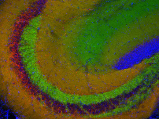
Two Synaptobrevin isoforms were identified in the mammalian CNS, synaptobrevin1 (VAMP1 or p18-1) and synaptobrevin2 (VAMP2 or p18-2) that differ in their regional distribution within the brain, indicating isoform-specific roles in neuroexocytosis (7).
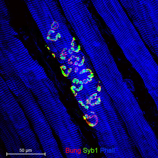
Synaptobrevin1 (VAMP1) is supposed to be essential for the maintenance of nerve impulse transmission in neuromuscular synapses. In addition, it is present on secretory granules of neuroendocrine cells. Synaptobrevin2 (VAMP2) is more abundant and widely distributed in the brain and has been shown to be mainly involved in the assembly of effective SNARE complexes, Ca2+-dependent SV exocytosis, and fast endocytosis in hippocampal synapses (8). It is also expressed in spinal cord dorsal horn neurons and implicated in inflammatory pain sensitization (9).
Synaptobrevins are target molecules for tetanus and several of the botulinal neurotoxins which cleave the protein at single sites in the C-terminal portion of the molecule and thereby disrupt neurotransmitter release (10).
Certificates
ISO 9001 2015 Quality Management System and Green Lab Platinum certification level for sustaining laboratory processes.


Newsletter
Sign up for our newsletter and get the latest updates and news.