Cat. No.: 101 102
Amount: 200 µl
Price:
$360.00
|
|

|
|
| Cat. No. 101 102 |
200 µl antiserum, lyophilized. For reconstitution add 200 µl H2O, then aliquot and store at -20°C until use. Antibodies should be stored at +4°C when still lyophilized. Do not freeze! |
| Applications | |
| Immunogen | Full-length native rat Synaptophysin (UniProt Id: P07825) |
| Reactivity |
Reacts with: mouse (Q62277), rat (P07825), human (P08247), Guinea pig, rabbit. Other species not tested yet. |
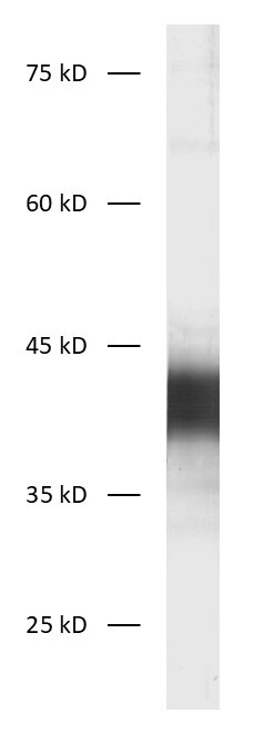
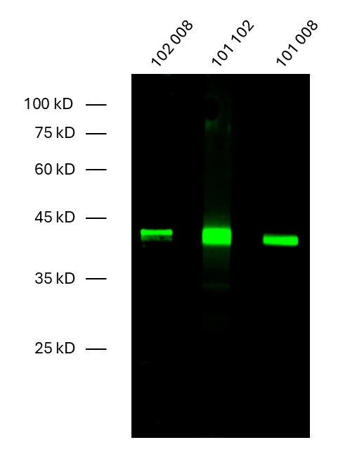
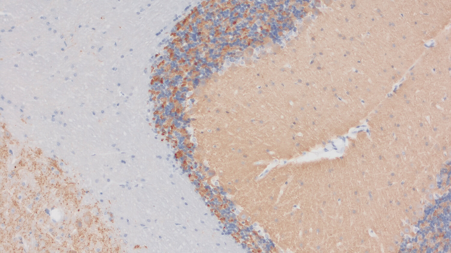
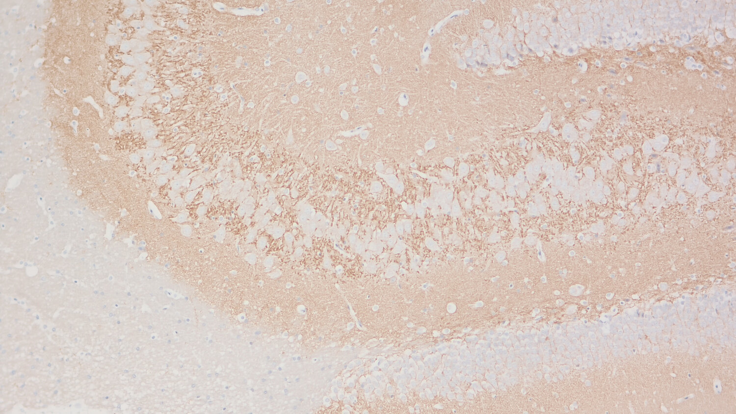
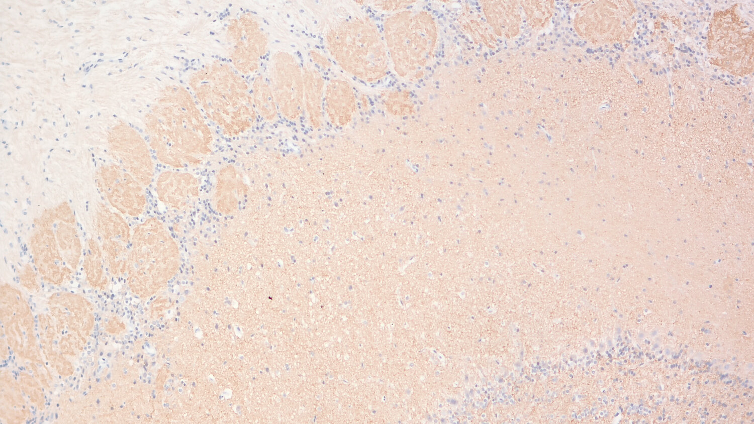
| Specificity | Recognizes Synaptophysin1 and 2 (Synaptoporin, p38-2). |
| Remarks |
ICC: The following fixatives are possible: 4% formaldehyde/PFA, methanol. |
| Data sheet | Datasheet 101_102 |
|
|
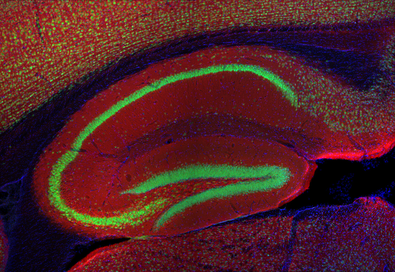
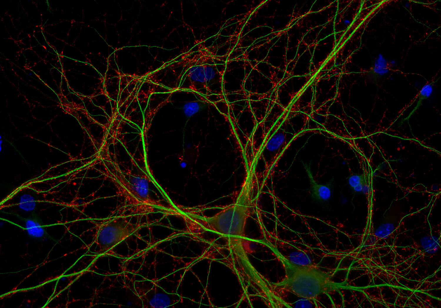
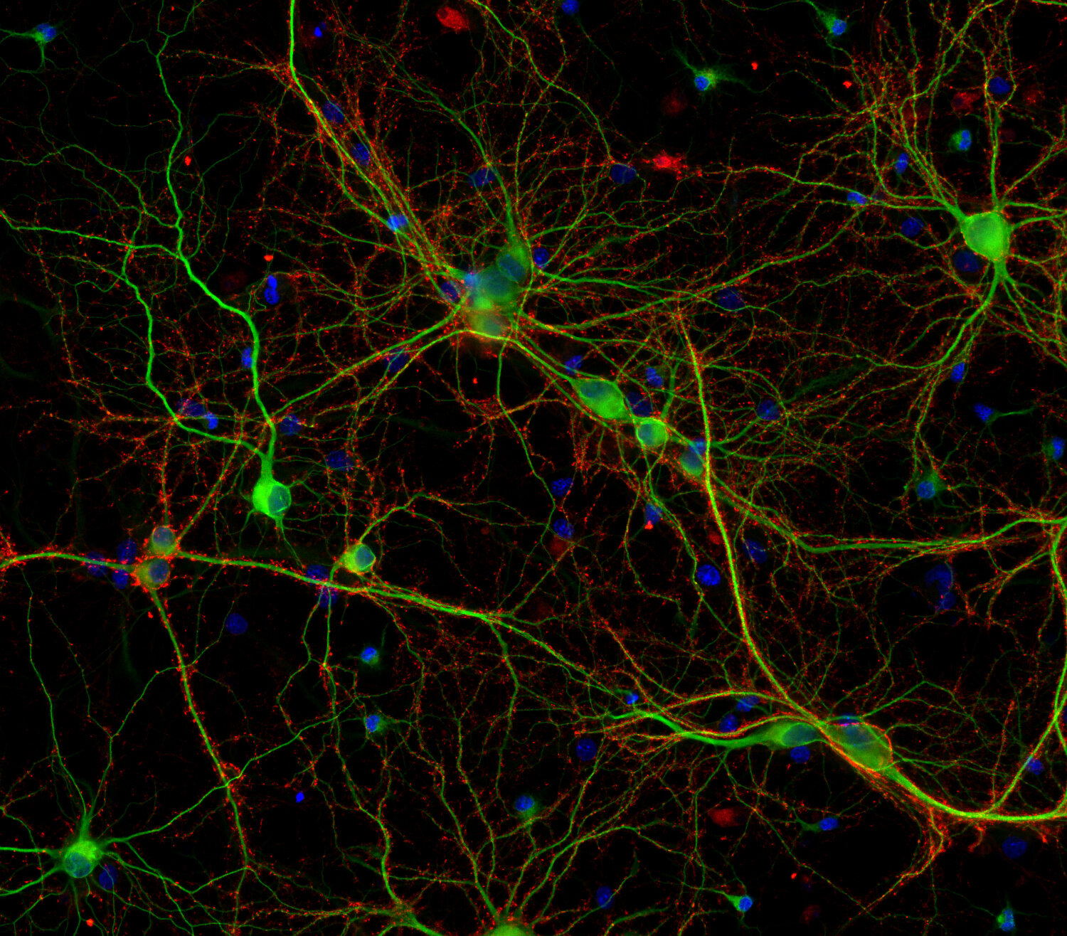
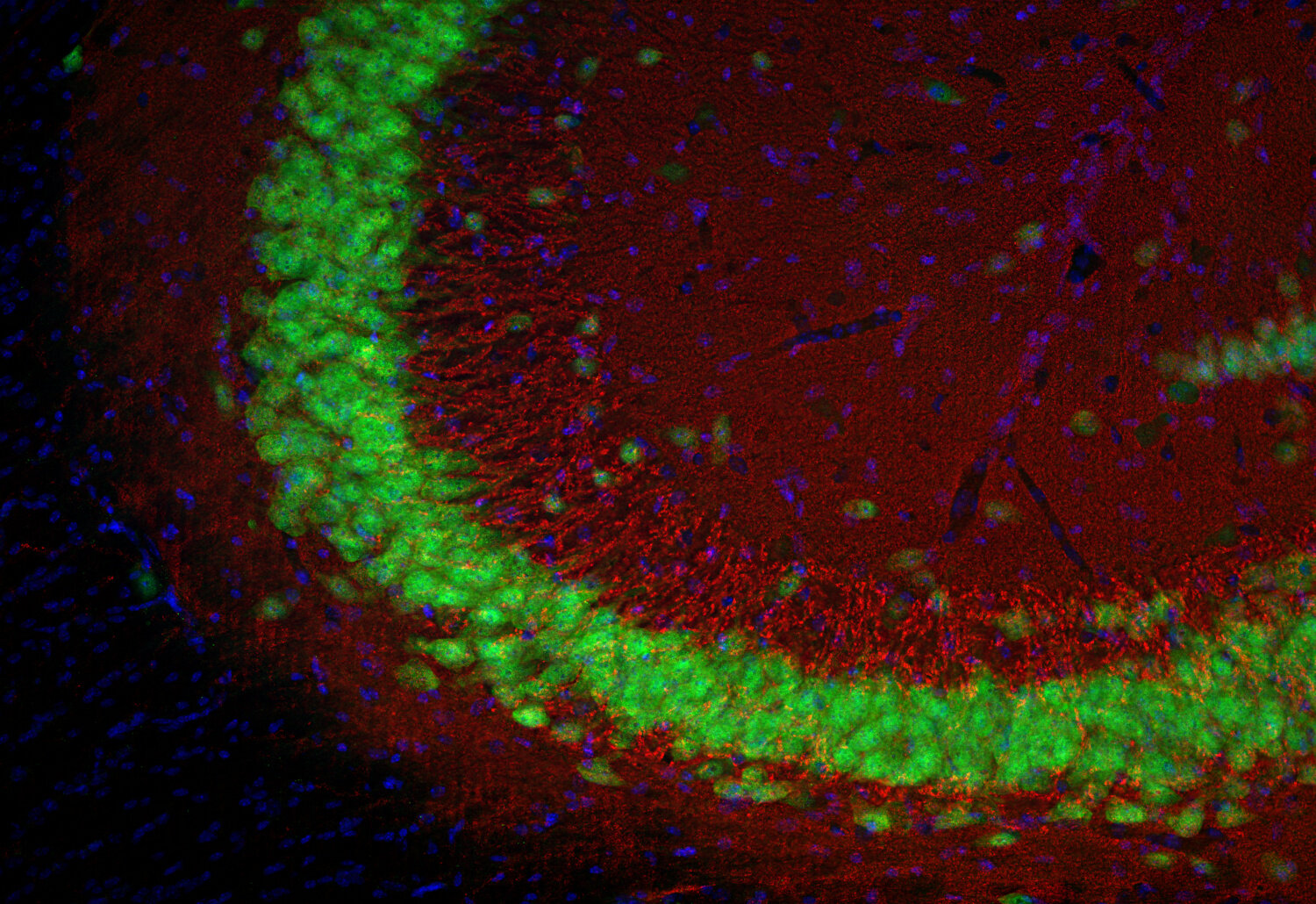
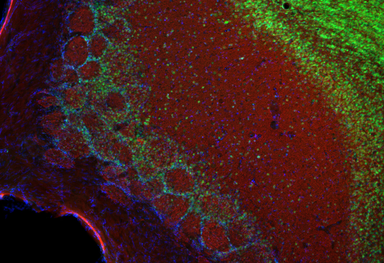
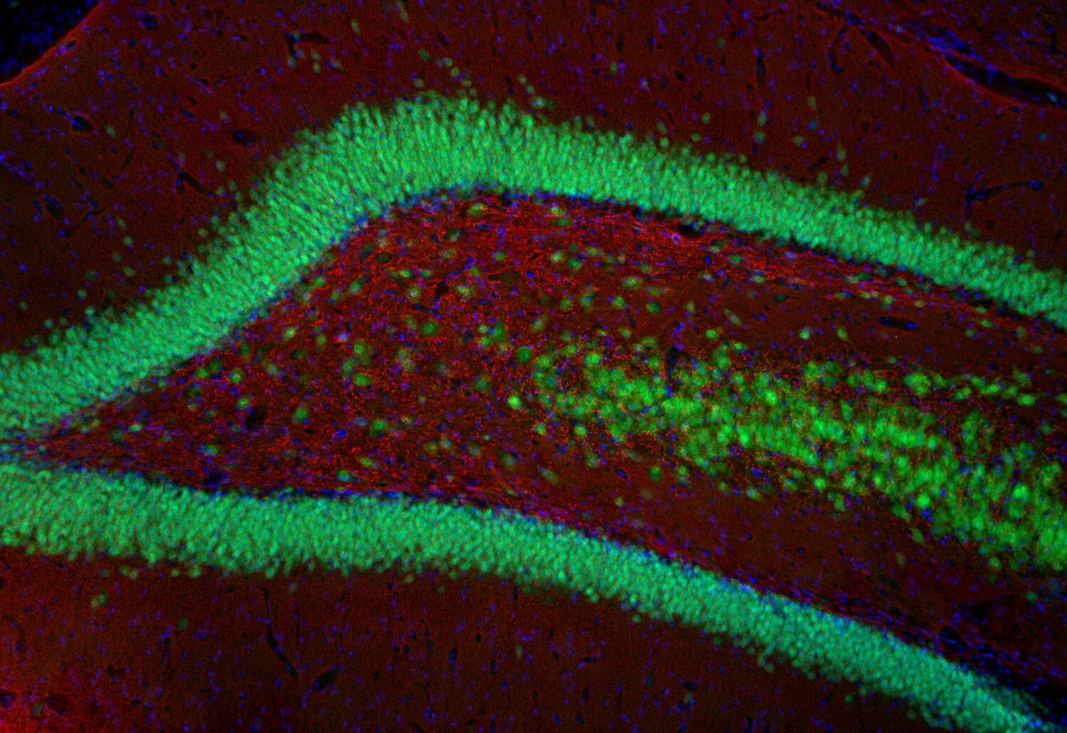
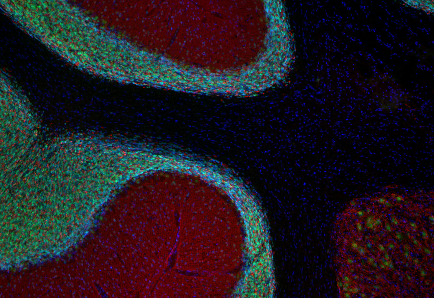
Synaptophysin1/2 (red) and NeuN (green) staining in mouse hippocampus
Synaptophysin1, also referred to as p38-1, is a membrane glycoprotein of synaptic vesicles that is ubiquitously expressed in all neurons and in many endocrine cells. It is currently the most widely used marker for nerve terminals and probably the best marker for the pathologist in differentiating neuroendocrine tumors.
Synaptophysin1 has four transmembrane domains with both N- and C-terminus facing the cytoplasm. It binds to synaptobrevin1 and synaptobrevin2 in detergent extracts but its function has not been elucidated completely. It forms a complex with dynamin at high Ca2+ concentration suggesting an involvement in synaptic vesicle endocytosis. As typical for synaptic vesicle proteins, synaptophysin1 represents a small protein family with two additonal members, synaptoporin and panthophysin.
Synaptoporin, also known as synaptophysin2 and p38-2, is highly homologous to synaptophysin1 but encoded by a different gene. Like synaptopysin1, synaptoporin contains four transmembrane regions and a short cytoplasmic tail. Unlike synaptophysin1, it is not glycosylated.
The distributions of synaptophysin1 and synaptoporin are different. Synaptophysin1 is more uniformly expressed whereas synaptoporin is particularly enriched in mossy fiber synapses in the hippocampus. It is thus an excellent marker for subsets of synapses.
Certificates
ISO 9001 2015 Quality Management System and Green Lab Platinum certification level for sustaining laboratory processes.


Newsletter
Sign up for our newsletter and get the latest updates and news.